| Journal of Endocrinology and Metabolism, ISSN 1923-2861 print, 1923-287X online, Open Access |
| Article copyright, the authors; Journal compilation copyright, J Endocrinol Metab and Elmer Press Inc |
| Journal website https://jem.elmerpub.com |
Original Article
Volume 16, Number 4, August 2026, pages 172-181

Corticotropin-Releasing Hormone Test for Diagnosing Central Adrenal Insufficiency: A Retrospective Cross-Sectional Study
Masashi Yoshikawaa  , Kazuhiko Horiguchia, e, Tetsuya Takamizawaa, b, Sho Sekiguchia, Yutaka Watanukia, Satoshi Yoshinoa, Shunichi Matsumotoa, Rei Yamaguchic, Masahiko Tosakac, d, Masanobu Yamadaa, Eijiro Yamadaa
aDivision of Endocrinology and Metabolism, Department of Internal Medicine, Gunma University Graduate School of Medicine, Maebashi 371-8511, Japan
bDepartment of Diabetes and Endocrinology, JA Nagano Koseiren Saku Medical Center, Nagano 384-0301, Japan
cDepartment of Neurosurgery, Gunma University Graduate School of Medicine, Maebashi 371-8511, Japan
dDepartment of Hypothalamic and Pituitary Surgery, Toranomon Hospital, Minato-ku, Tokyo 105-8470, Japan
eCorresponding Author: Kazuhiko Horiguchi, Division of Endocrinology and Metabolism, Department of Internal Medicine, Gunma University Graduate School of Medicine, Maebashi, Gunma 371-8511, Japan
Manuscript submitted April 23, 2026, accepted June 17, 2026, published online August 24, 2026
Short title: CRH Test for Central Adrenal Insufficiency
doi: https://doi.org/10.14740/jem1650
| Abstract | ▴Top |
Background: The diagnostic cutoff for central adrenal insufficiency (AI) is traditionally set at a peak cortisol level of 18 µg/dL during stimulation tests. However, modern high-specificity cortisol assays typically yield lower values, necessitating a re-evaluation of diagnostic thresholds. This study aimed to determine the optimal cutoff values and to explore a practical diagnostic approach for the corticotropin-releasing hormone (CRH) stimulation test using a contemporary assay system.
Methods: We retrospectively analyzed 83 patients with hypothalamic-pituitary structural lesions who underwent a CRH test between 2016 and 2020. Patients were classified into an adrenal sufficiency group (AS, n = 44) or an AI group (n = 39) based on comprehensive clinical assessment, including symptoms and long-term follow-up. Receiver operating characteristic (ROC) curve analysis was performed to identify diagnostic thresholds.
Results: Basal and peak cortisol levels were significantly lower in the AI group than in the AS group (P < 0.05). ROC analysis for diagnosing AI showed that peak serum cortisol had the highest diagnostic performance (area under the curve, 0.94). The optimal peak cortisol cutoff value was 16.3 µg/dL (sensitivity: 94.9%, specificity: 79.6%). A peak cortisol threshold of 17.0 µg/dL achieved 100% sensitivity, whereas a basal cortisol level of 3.3 µg/dL, a peak cortisol threshold of 10.0 µg/dL, and a peak adrenocorticotropic hormone (ACTH) level of 36.5 pg/mL provided 100% specificity. Notably, peak cortisol values in the 10.1–16.9 µg/dL range showed significant overlap between the groups. Delayed ACTH peaks (≥ 60 min) were more frequent in the AI group but lacked definitive diagnostic specificity.
Conclusions: By integrating our findings with contemporary literature using modern high-specificity assays, we propose a practical step-by-step clinical approach that sequentially incorporates a basal cortisol rule-in threshold (≤ 3.0 µg/dL), peak ACTH threshold (≤ 35 pg/mL), and a screening threshold for peak cortisol (17.0 µg/dL). Clinical judgment remains essential for patients with peak cortisol values between 10.1 and 16.9 µg/dL, for whom diagnosis and management should be individualized based on clinical symptoms and biochemical findings rather than a single binary threshold.
Keywords: Adrenal insufficiency; Cortisol; Corticotropin-releasing hormone test; Hypothalamus; Pituitary
| Introduction | ▴Top |
Adrenocorticotropic hormone (ACTH) deficiency due to pituitary and hypothalamic diseases causes central adrenal insufficiency (AI), with clinical manifestations ranging from asymptomatic cases to overt adrenal failure. Even asymptomatic individuals may develop hormone deficiency under stress, which occasionally progresses to adrenal crisis. Therefore, appropriate hormone replacement therapy is necessary.
Several stimulation tests have been used to evaluate the hypothalamic–pituitary–adrenal (HPA) axis. The Endocrine Society guidelines recommend the insulin tolerance test (ITT) and rapid ACTH test [1], while in Japan, the corticotropin-releasing hormone (CRH) test is additionally recommended [2]. In both guidelines, the peak cortisol cutoff value obtained from these tests is set at 18 µg/dL (500 nmol/L). However, this cutoff value was established using assay kits with low cortisol specificity; with the currently used high-specificity assay systems, the appropriate cutoff value is expected to be lower than 18 µg/dL (500 nmol/L). Comparative analysis of rapid ACTH stimulation test results indicated that current assay kits have reduced the peak cortisol cutoff value from 18 µg/dL to approximately 14–16 µg/dL [3–5].
The ITT has traditionally been regarded as the gold standard for evaluating the HPA axis [6]. Adequate stimulation during the ITT requires lowering the blood glucose level to 40–45 mg/dL; however, the test is contraindicated in patients with ischemic heart disease or epilepsy. Even when feasible, the ITT imposes a substantial burden on patients and requires close monitoring by medical staff. While the rapid ACTH test is safer by comparison, cortisol secretion from the adrenal cortex may show a normal response if the duration since the onset of pituitary ACTH impairment is short; thus, it is unsuitable for acute or short-duration cases.
By contrast, the CRH test has fewer adverse effects and is therefore safer; furthermore, it can be performed simultaneously with the thyrotropin-releasing hormone (TRH) and gonadotropin-releasing hormone tests. Consequently, it offers the advantage of being safely administered with minimal patient burden, regardless of the timing of onset, in cases where hypopituitarism is suspected. Given these factors, in Japanese clinical practice, many suspected cases of hypopituitarism are evaluated using the CRH test alone. In interpreting these results, endocrinologists determine the necessity of hydrocortisone replacement by comprehensively evaluating individual clinical and laboratory findings while considering the 18-µg/dL threshold stipulated in the aforementioned guidelines and the fact that modern assay systems tend to yield lower values. However, to date, few studies have validated the precise cutoff values and clinical utility of the CRH test using contemporary assay systems. Therefore, the present study aimed to evaluate the utility of the CRH test for central AI in the context of current assay systems.
| Materials and Methods | ▴Top |
Study design and population
This retrospective cross-sectional study included consecutive patients who underwent a CRH stimulation test at Gunma University Hospital between 2016 and 2020 and presented with structural abnormalities of the pituitary or hypothalamus on imaging. Patients were excluded if they had insufficient data, markedly elevated baseline cortisol levels, and functional pituitary adenomas. Furthermore, none of the patients had a prior history of corticosteroid treatment at pharmacological doses. The predefined inclusion and exclusion criteria are summarized in Table 1. The final sample size was determined by the total number of eligible patients who met the predefined inclusion and exclusion criteria during the study period.
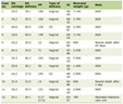 Click to view | Table 1. Eligibility Criteria for the Study Population |
The reference standard for the diagnosis of central AI was established based on expert clinical judgment, which comprised a comprehensive evaluation of clinical symptoms, basal pituitary-adrenal laboratory findings, and the clinical decision to initiate hydrocortisone replacement therapy. This clinical judgment was made collectively by endocrinologists at our hospital in patients presenting with symptoms suggestive of AI, such as nausea, malaise, anorexia, or hyponatremia, alongside the status of other pituitary hormone axes. These assessments were made considering the guideline-defined peak cortisol threshold of 18 µg/dL while also considering that equivalent values in modern assays typically range from 14 to 16 µg/dL. Even in cases where regular hydrocortisone replacement was deemed unnecessary, the potential risk of AI was thoroughly explained to the patients. These individuals were provided with as-needed hydrocortisone prescriptions and were instructed to notify their physicians if they were to undergo invasive procedures or examinations. Patients continuing treatment at our hospital were followed until December 2024, while those referred to other institutions were followed until their final visit to our department.
Stimulation tests
The CRH stimulation test was performed in the early morning after an overnight fast and at least 30 min of bed rest. In patients who had been receiving hydrocortisone, treatment was discontinued at least 24 h before testing.
Synthetic CRH (100 µg; corticorelin, Nipro ES Pharma Corporation, Osaka, Japan) was administered intravenously as a bolus. Blood samples were collected at baseline and at 30, 60, and 120 min after CRH administration to measure serum ACTH and cortisol levels. Peak ACTH and cortisol values were defined as the highest concentrations observed after CRH administration.
To describe variability in ACTH response patterns, an exploratory operational definition was applied: a peak ACTH concentration occurring at ≥ 60 min after CRH administration was defined as a delayed peak. This definition was adopted for descriptive purposes and was informed by the European Thyroid Association guideline, which describes a delayed thyroid-stimulating hormone peak (≥ 60 min) during the TRH stimulation test as a supportive finding in central hypothyroidism [7].
Hormonal assay
Serum ACTH levels were measured using an Elecsys ACTH electrochemiluminescence immunoassay (Roche Diagnostics, Indianapolis, IN, USA), and cortisol levels were measured using the Elecsys Cortisol II, electrochemiluminescence immunoassay (Roche Diagnostics). Reference ranges for ACTH and cortisol were 7.2–63.3 pg/mL and 3.0–19.6 µg/dL (82.8–540 nmol/L), respectively. All additional biochemical parameters were analyzed using standard laboratory methods.
Ethical compliance with human study
This study was conducted in compliance with the ethical standards of the responsible institution on human subjects as well as with the Declaration of Helsinki and was approved by the Ethics Committee on Human Research of Gunma University (Approval Number: HS2019-082).
Statistical analysis
To address potential sources of bias, selection bias was minimized by including all consecutive patients who met the predefined criteria, and measurement bias was mitigated through the use of a standardized single-assay system (Elecsys Cortisol II) for all participants. Hormone level data from the clinical study are presented as median and interquartile range. Proportions were compared using the Pearson Chi-square test, and continuous variables between the two groups were compared using the non-parametric Wilcoxon test because the data were not normally distributed. The Fisher’s exact test was used for contingency table analyses between the two groups.
Receiver operating characteristic (ROC) curve analysis was performed, and the optimal cutoff values were determined by the Youden index, considering sensitivity and specificity. The 95% confidence intervals (CIs) for the ROC curves were calculated using R (R Core Team, Vienna, Austria). All other statistical analyses were performed using JMP Pro (version 12.0.1; SAS, Cary, NC, USA), and statistical significance was defined as P < 0.05. This study was prepared and reported in accordance with the Standards for Reporting Diagnostic Accuracy Studies 2015 (STARD 2015) guidelines.
| Results | ▴Top |
Study population and baseline characteristics of the AS and AI groups
The study flowchart is shown in Figure 1. Of the 156 patients who underwent the CRH test, we excluded patients with insufficient data (n = 26), markedly elevated baseline cortisol levels (n = 5), and functional pituitary adenomas (n = 36). Subsequently, 89 patients remained. Among them, 44 patients had never received hydrocortisone prior to the CRH test or through the end of the follow-up period and were classified as the adrenal sufficiency (AS) group. The remaining 45 patients received hydrocortisone either before (n = 40) or immediately after (n = 5) the CRH test. During the follow-up, hydrocortisone was discontinued in six patients, who were further excluded. Then, the remaining 39 patients were classified as the AI group. The underlying etiologies in the AS group were lymphocytic hypophysitis in three patients, non-functioning pituitary adenoma in 29 patients, Rathke cleft cyst in nine patients, craniopharyngioma in two patients, and surgery for a Rathke cleft cyst in one patient. In the AI group, the etiologies included lymphocytic hypophysitis in four patients, immunoglobulin G4-related hypophysitis in one patient, pituitary apoplexy in one patient, non-functioning pituitary adenoma in 20 patients, Rathke cleft cyst in two patients, craniopharyngioma in three patients, and postoperative status in eight patients (non-functioning pituitary adenoma in two, Rathke cleft cyst in one, craniopharyngioma in two, functioning tumors in two, and other causes in one ).
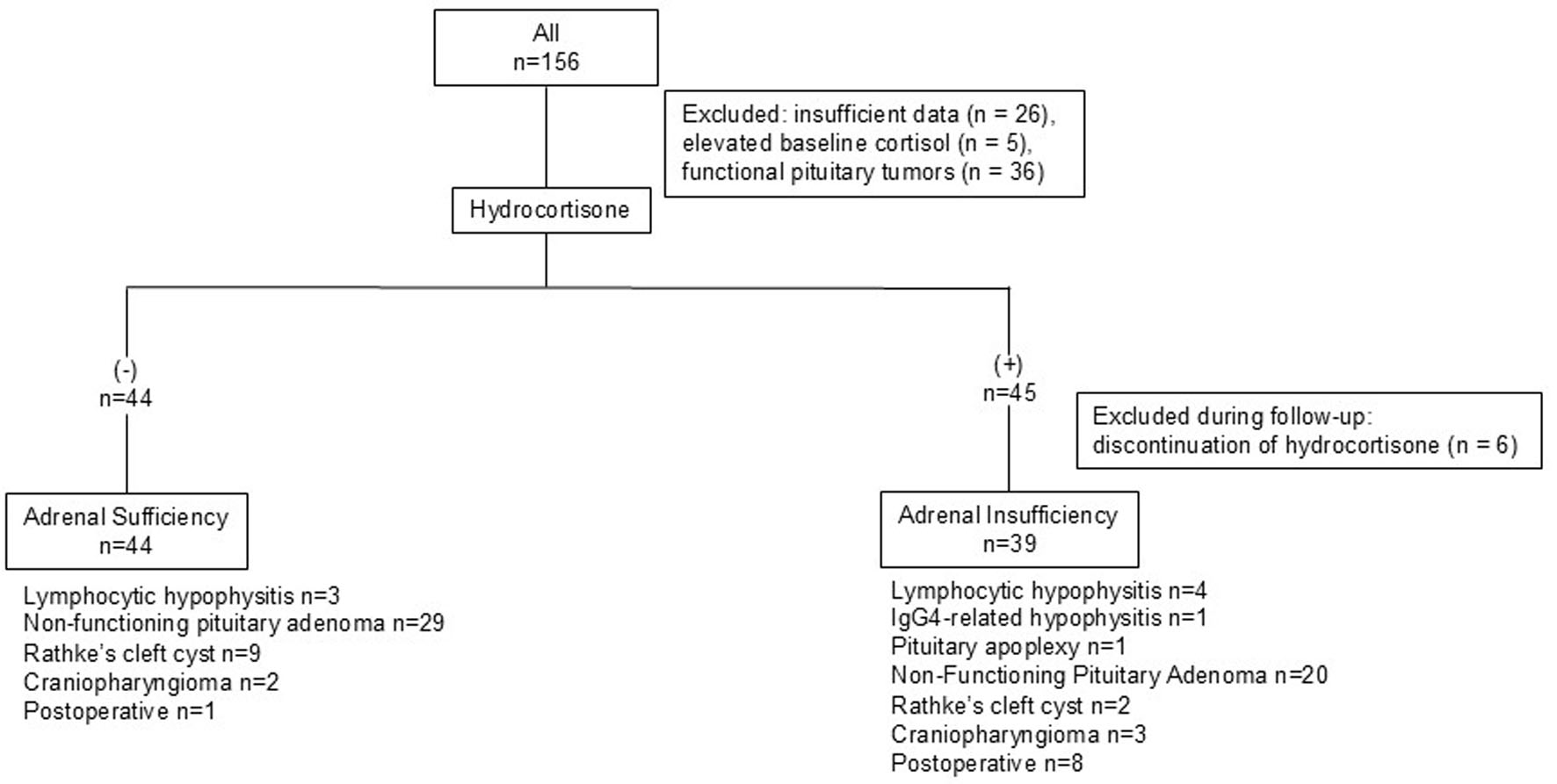 Click for large image | Figure 1. Study design. Flow diagram of patient selection and etiological classification. Among 156 patients, those with insufficient data, elevated baseline cortisol, or functional pituitary tumors were excluded. Then, patients were stratified according to hydrocortisone use (−, n = 44; +, n = 45), with additional exclusions for discontinuation during follow-up. The final cohorts comprised the adrenal sufficiency group (hydrocortisone −, n = 44) and the adrenal insufficiency group (hydrocortisone +, n = 39), along with their underlying etiologies. |
The baseline characteristics are presented in Table 2. The AS and AI groups included 44 and 39 patients, respectively. The respective median ages and body weight (BW) were not significantly different between the groups. Severe adult growth hormone deficiency (AGHD) occurred in 18 (40.9%) and 29 (74.3%) patients in the AS and AI groups, respectively. Hypogonadotropic hypogonadism occurred in 17 (38.6%) and 30 (76.9%) patients in the AS and AI groups, respectively. Central hypothyroidism occurred in 0 (0%) and 22 (56.4%) patients in the AS and AI groups, respectively.
Click to view | Table 2. Baseline Characteristics of the AS and AI Groups |
Clinical manifestations in the AI group included nausea in three patients, fatigue in 20 patients, loss of appetite in eight patients, hyponatremia in five patients, and absence of symptoms in 15 patients. Some patients presented with multiple symptoms.
Basal hormone levels and ACTH–cortisol responses to the CRH test
Pre-ACTH levels were significantly lower in the AI group than in the AS group (Fig. 2a). Similarly, pre-cortisol levels were significantly lower in the AI group than in the AS group (Fig. 2b). Peak ACTH responses to the CRH test showed significantly lower peaks in the AI group than in the AS group (Fig. 2c). Peak cortisol levels were also significantly lower in the AI group than in the AS group (Fig. 2d).
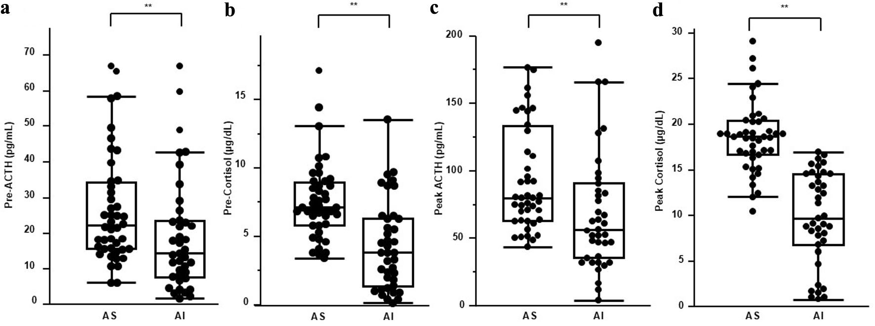 Click for large image | Figure 2. Baseline and peak CRH-stimulated ACTH and cortisol levels. Comparison of basal and CRH-stimulated hormone levels between patients with AS and those with AI. (a) Baseline plasma ACTH levels (pre-ACTH), (b) baseline serum cortisol levels (pre-cortisol), (c) peak ACTH levels (peak ACTH), and (d) peak serum cortisol levels (peak cortisol) during the CRH stimulation test are shown for the AS and AI groups. Box plots show the median (horizontal line), interquartile range (box), whiskers, and individual data points. Baseline and peak hormone levels are significantly lower in the AI group than in the AS group (P < 0.01). For optimal visualization of the primary data distributions, extreme outliers exceeding 200 pg/mL for peak ACTH in panel (c) and 30 µg/dL for peak cortisol in panel (d) were omitted from the plots; however, all data points were fully included in the statistical analyses. ACTH: adrenocorticotropic hormone; AI: adrenal insufficiency; AS: adrenal sufficiency; CRH: corticotropin-releasing hormone. |
Delayed response of ACTH on the CRH test (Elecsys Cortisol II)
We also examined delayed ACTH after CRH stimulation (Table 3). Delayed ACTH occurred more frequently in the AI group than in the AS group (59%, 23/39 versus 32%, 14/44; odds ratio = 3.08, 95% CI: 1.25–7.57; Fisher’s exact test, two-sided P = 0.016).
Click to view | Table 3. Delayed Response of ACTH in the CRH Test |
Cutoff values for pre-stimulation ACTH, peak ACTH, pre-stimulation cortisol, and peak cortisol on the CRH test for diagnosing AI
ROC analysis showed that pre-stimulation ACTH had an area under the curve (AUC) of 0.67 (95% CI: 0.55–0.79), and the optimal cutoff value determined by the Youden index was 12.1 pg/mL (sensitivity, 43.5%; specificity, 90.9%) (Fig. 3a). Similarly, pre-stimulation serum cortisol demonstrated an AUC of 0.79 (95% CI: 0.69–0.90), with an optimal cutoff value of 5.6 µg/dL (sensitivity, 71.8%; specificity, 79.6%). A lower cutoff of 3.3 µg/dL achieved 100% specificity, although sensitivity decreased to 46.1% (Fig. 3b). For peak ACTH, the AUC was 0.70 (95% CI: 0.58–0.82), and the optimal cutoff value was 62.6 pg/mL (sensitivity, 56.4%; specificity, 81.8%). A lower cutoff of 36.5 pg/mL achieved 100% specificity, although the sensitivity decreased to 28.2% (Fig. 3c). Finally, peak serum cortisol exhibited the highest diagnostic performance with an AUC of 0.94 (95% CI: 0.89–0.98). The optimal cutoff value was 16.3 µg/dL, corresponding to a sensitivity of 94.9% and a specificity of 79.6%. A higher cutoff of 17.0 µg/dL achieved 100% sensitivity, with a specificity of 70.5%, and a lower cutoff value of 10.0 µg/dL achieved 53.8% sensitivity, with a specificity of 100% (Fig. 3d). Among these parameters, peak serum cortisol demonstrated the highest diagnostic accuracy.
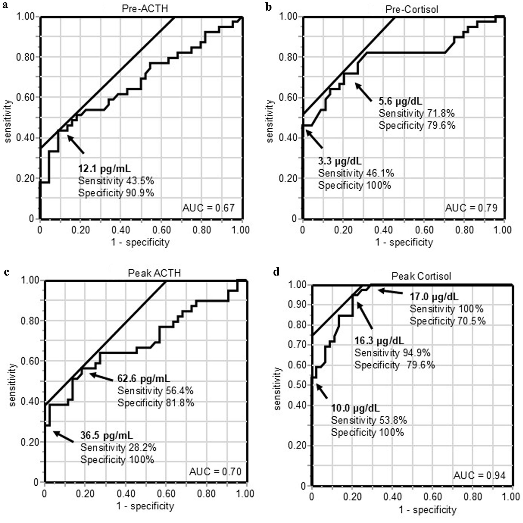 Click for large image | Figure 3. ROC curves for baseline and CRH-stimulated peak ACTH and cortisol levels in diagnosing central adrenal insufficiency. ROC curves for CRH test parameters in the diagnosis of adrenal insufficiency. (a) Pre-ACTH, (b) pre-cortisol, (c) peak ACTH, and (d) peak cortisol levels. The AUC is shown in each panel. Arrows indicate optimal cutoff values determined by the Youden index; alternative cutoff values providing 100% sensitivity or specificity are also indicated where applicable. ACTH: adrenocorticotropic hormone; AUC: area under the curve; CRH: corticotropin-releasing hormone; ROC: receiver operating characteristic. |
| Discussion | ▴Top |
This study examined the usefulness of the CRH test for diagnosing central AI in clinical practice at a single institution using recent assay systems, and the findings suggest that the peak cortisol value has diagnostic relevance. Similar to this study, Izuchi et al evaluated peak cortisol levels during CRH stimulation tests for the diagnosis of AI using the Elecsys Cortisol II assay. In their study, the cutoff value for cortisol 30 min after stimulation was 12.6 µg/dL [8]. Their study reported cortisol levels of 16.2 (14.3–17.9) µg/dL in the normal adrenal function group and 1.7 (0.6–8.6) µg/dL in the AI group. Compared with our study, where the cortisol levels were 18.7 (16.8–20.5) µg/dL in the AS group and 9.8 (6.9–14.6) µg/dL in the AI group, the cortisol values in the AI group of Izuchi et al’s study were markedly lower.
Upon reviewing the breakdown of diseases, in Izuchi et als study, immune checkpoint inhibitor (ICI)-related pituitary dysfunction accounted for more than half of the AI group (51 out of 91 cases). ICI-related pituitary dysfunction is characterized by severe ACTH deficiency, often reaching near-total depletion. By contrast, in our study, among the 39 cases of AI, inflammatory diseases accounted for only five cases (including lymphocytic hypophysitis combined with IgG4-related hypophysitis), while 34 cases involved pituitary space-occupying lesions (including one case of pituitary apoplexy) pre- or post-surgery. Therefore, it is considered that our study included many cases of partial ACTH deficiency caused by tumor compression. This difference in the selected disease populations likely led to the variations in peak cortisol levels (or 30-min values) and the resulting cutoff values in the AI group.
Furthermore, Mitsui et al conducted a similar study using conventional and modern assays, reporting cutoff values for diagnosis of 12.6 µg/dL for the 30-min cortisol value and 14.3 µg/dL for the peak cortisol value [9]. Despite including cases measured with conventional assays, their cutoff values remained lower than those in our study. An examination of the disease breakdown in their study revealed that among 87 cases of AI, isolated ACTH deficiency, characterized by severe, near-total depletion of ACTH, accounted for approximately 30% (32 cases). These findings suggest that cutoff values may be substantially influenced not only by the generation of the assay used but also by the characteristics of the selected disease groups.
The status of the ITT as the gold standard, as well as the adoption of 18 µg/dL as the diagnostic cutoff value for central AI, are rooted in clinical studies conducted in healthy individuals between the 1970s and early 1990s. The ITT is considered an HPA axis test because it mimics such stress conditions, and Allen et al and Nelson et al noted a peak cortisol level of 15 µg/dL in healthy individuals [10, 11]. Similarly, Streeten et al reported that healthy participants reached a cortisol level of 29.4 ± 2.0 µg/dL at 60 min during the ITT [6]. Bliss et al measured 17-hydroxycorticosteroids during the ITT and described peak values of 25–34 µg/dL [12]. Four studies assessing peak 11-hydroxycorticosteroids levels during the ITT showed that minimum values were consistently ≥ 21 µg/dL [13–16]. Similarly, Staub et al also reported that the minimum peak cortisol level was 18.9 µg/dL [17]. These results indicate that an intact HPA axis should yield peak cortisol levels ≥ 18–20 µg/dL during the ITT. Based on the actual data from these reports, the peak value of 18.0 µg/dL should be interpreted as a threshold for ensuring 100% diagnostic sensitivity rather than as a definitive diagnostic cutoff value. In clinical practice, this traditional threshold is largely based on the principle that missing a diagnosis of AI because of false negatives poses critical, life-threatening risks to patients. In the present study, 100% sensitivity was observed at 17.0 µg/dL, whereas the statistically optimal diagnostic cutoff value was 16.3 µg/dL. Notably, the study by Izuchi et al did not report a specific threshold value achieving 100% sensitivity [8]. Meanwhile, Mitsui et al reported a 100% sensitivity threshold of 17.1 µg/dL [9]. Given that Mitsui et al’s study utilized a mixture of conventional and modern assays, their reported threshold value would theoretically be lower if evaluated exclusively with contemporary high-specificity assays. Taking these factors into comprehensive consideration, our findings support the use of 17.0 µg/dL as a practical screening threshold for peak cortisol levels in the CRH test to optimize patient safety in real-world clinical settings.
Several reports have also discussed the relationship between the ITT and CRH test. In patients receiving long-term exogenous glucocorticoids, the peak cortisol level was lower in the CRH test than in the ITT test, although they remained correlated, and both were useful for diagnosing central AI [18]. Conversely, in 2003, Schmidt et al reported that when the ITT result was used as the reference, the CRH test result maximized sensitivity and specificity at a peak cortisol level of 13.7 µg/dL; however, sensitivity was only 75% and specificity was 98%, resulting in a high false negative. Thus, the CRH test was considered to have limited diagnostic utility [19]. Maghnie et al evaluated adrenal cortical function in cases of severe adult growth hormone deficiency. Using a cortisol peak of 18 µg/dL as the cutoff value, seven out of 12 patients were diagnosed with AI via the ITT. Of the seven cases of AI identified by the ITT, only three were able to be diagnosed using the CRH test. This suggests that the same cutoff value may not be applicable, with inconsistency in CRH responsiveness proposed as a contributing factor [20]. Dullaart et al reported that healthy individuals show a stronger cortisol response to the CRH test than to the ITT, whereas individuals with AI show a weaker response to the CRH test than to the ITT [21]. Based on these reports, the cortisol response may be higher in the CRH test than in the ITT. Therefore, we must recognize that when diagnosing AI using only the CRH test, there is a risk of missing cases requiring supplementation due to false negatives.
Additionally, as is also true when performing the ITT, as long as a cutoff value is determined within a continuous range of numerical data, clinical judgment will always be required for interpreting values near that threshold. In the present study, a peak cortisol level of 17.0 µg/dL was the value that ensured 100% diagnostic sensitivity for AI; however, the AS and AI groups overlapped within the range of 10.1–16.9 µg/dL. Similarly, Mitsui et al reported that a peak cortisol level of 17.1 µg/dL provided 100% diagnostic sensitivity, yet the AS and AI groups coexisted within the range of 10.0–17.0 µg/dL [9]. For cases where CRH test peak values fall within these ranges, Mitsui et al proposed that the necessity of hydrocortisone replacement should be determined by referencing basal DHEA-S levels [9]. Likewise, in the present study, the normal adrenal function and AI groups overlapped in cases where peak cortisol levels were 10.1–16.9 µg/dL, requiring comprehensive clinical judgment based on symptoms and laboratory findings. We hypothesized that a delayed peak ACTH response might serve as a useful reference in such cases; however, in practice, delayed ACTH peak responses were significantly more frequent in the AI group than in the AS group, although a substantial proportion of delayed responses was also observed in the AS group. Therefore, delayed peak ACTH levels cannot be considered a useful parameter for establishing a definitive diagnosis. Consequently, managing patients with peak cortisol values within the 10.1–16.9 µg/dL range remains a clinical challenge that cannot be resolved by a single binary threshold. In real-world practice, these patients require individualized, case-by-case management strategies. If clinicians elect to initiate hydrocortisone replacement because of significant symptoms, careful monitoring for iatrogenic Cushing’s syndrome is warranted. Conversely, if replacement is deferred, close vigilance for the development of AI symptoms or adrenal crisis remains essential.
Basal cortisol levels have long been proposed as a useful tool for the diagnosis of AI. Schmidt et al reported that when the ITT is used as the reference, a basal cortisol level of 3.55 µg/dL (98 nmol/L) demonstrated 100% specificity, whereas 10.32 µg/dL (285 nmol/L) demonstrated 100% sensitivity [19]. Historically, the landmark meta-analysis by Kazlauskaite et al, which reviewed studies using older-generation cortisol assays, reported a weighted mean “rule-in” threshold of ≤ 5.0 µg/dL, though several strict cohorts within that analysis frequently identified lower definitive thresholds ranging between 3.0 and 3.5 µg/dL [22]. Given that older assays generally exhibited greater cross-reactivity and produced higher absolute values than contemporary platforms, these historical thresholds would be expected to shift lower in modern assay systems. In fact, a recent investigation by de Vries et al utilizing the modern, high-specificity Elecsys Cortisol II assay with the ITT reference demonstrated that a basal cortisol cutoff of 82 nmol/L—which precisely converts to 2.97 µg/dL (approximately 3.0 µg/dL)—corresponded to 100% specificity for diagnosing central AI [23]. In our current study, which also employed the Elecsys Cortisol II assay, a basal cortisol level of 3.3 µg/dL yielded 100% specificity. Integrating our statistical findings with the contemporary literature (2.97 µg/dL) [23] and prioritizing day-to-day clinical practicality, our findings support ≤ 3.0 µg/dL as a practical rule-in threshold to safely omit stimulation tests and initiate hydrocortisone replacement.
Basal ACTH levels showed wide interindividual variation in the AS and AI groups, preventing construction of a meaningful ROC curve. This variability is attributed to the secretion of ACTH with low biological activity in cases of central AI [24, 25]. ACTH peak levels were distributed across a wide range above basal values, suggesting contributions from biologically low ACTH, and in some hypothalamic dysfunction cases, exaggerated responses [26, 27]. Nevertheless, we hypothesized that a distinctly low peak ACTH level—representing severe, near-total depletion of ACTH—could still hold substantial diagnostic relevance for central AI. Although the statistically optimal cutoff value of 62.6 pg/mL offered limited diagnostic utility due to modest accuracy (sensitivity, 56.4%; specificity, 81.8%), a detailed ROC table analysis successfully identified 36.5 pg/mL as a threshold that showed 100% specificity (0% false-positive rate) in our cohort for diagnosing central AI. To our knowledge, no prior studies have specifically evaluated or defined peak ACTH thresholds aimed at maximizing diagnostic specificity in the CRH test. While our actual statistical finding was 36.5 pg/mL, we propose a rounded, integer-based threshold of 35 pg/mL as a practical clinical rule-in criterion to enhance day-to-day clinical utility. Considering the fundamental physiological sequence of the HPA axis—where CRH stimulation triggers pituitary ACTH secretion, which subsequently drives adrenal cortisol output—employing a step-by-step diagnostic workflow based on this upstream-to-downstream cascade appears biologically plausible. Clinically, patients with a peak ACTH level ≤ 35 pg/mL were consistently classified as having central AI in our cohort. Conversely, for patients with a peak ACTH level exceeding 35 pg/mL, a higher numerical value does not guarantee adequacy; as noted above, circulating molecules may lack the biological potency required to stimulate the adrenal cortex. Therefore, in these upstream-sufficient or exaggerated cases, evaluating the final downstream adrenal response via peak cortisol levels (utilizing the aforementioned screening threshold of 17.0 µg/dL) may represent a physiologically plausible clinical approach to establishing an accurate diagnosis.
This study has some limitations. It had a retrospective single-center study design, and the number of cases was limited. Additionally, as symptoms of AI may also occur in other disorders, some patients classified as having AI may not have had true AI. Furthermore, the AS and AI groups included patients with impaired secretion of other pituitary hormones. In cases complicated by untreated growth hormone deficiency or hypothyroidism, delayed cortisol metabolism may result in peak responses to CRH stimulation being overestimated (i.e., appearing higher) relative to the patient’s true adrenal reserve. Although the ITT remains the gold standard for diagnosing central AI, this study was not designed to evaluate the diagnostic utility of the CRH stimulation test using the ITT as the reference standard. Lastly, the study population was predominantly Japanese, potentially limiting generalizability of the results.
In conclusion, this single-center retrospective study provides updated diagnostic insights for the CRH stimulation test using a contemporary, high-specificity cortisol assay. While the ITT remains the gold standard, the CRH test offers a safer, more convenient alternative, particularly for patients with contraindications or those requiring a less burdensome evaluation. Our findings suggest that while a basal serum cortisol level ≤ 3.0 µg/dL showed 100% specificity in our cohort to rule in central AI and may allow stimulation testing to be omitted, a single peak cortisol cutoff—such as the statistically optimal 16.3 µg/dL—is insufficient to manage the clinical complexities of the disease. We propose a practical step-by-step clinical approach (Fig. 4) that sequentially integrates basal cortisol (≤ 3.0 µg/dL), peak ACTH (≤ 35 pg/mL), and a conservative clinical screening threshold for peak cortisol (17.0 µg/dL). Patients with peak cortisol values between 10.1 and 16.9 µg/dL require individualized clinical assessment and management rather than a uniform diagnostic decision. If hydrocortisone replacement is initiated based on clinical symptoms, monitoring for overtreatment is warranted; conversely, if replacement is deferred, careful follow-up for the development of AI remains important. Further validation in independent studies utilizing contemporary, high-specificity assay systems across diverse patient populations—reflecting variations in underlying etiologies and degrees of pituitary longitudinal dysfunction—is needed to build upon our findings. Continuous clinical efforts, including our ongoing evaluations, will be essential to accumulate real-world evidence and further improve the clinical assessment of patients with peak cortisol values in the overlapping range, thereby supporting safer and more individualized patient management.
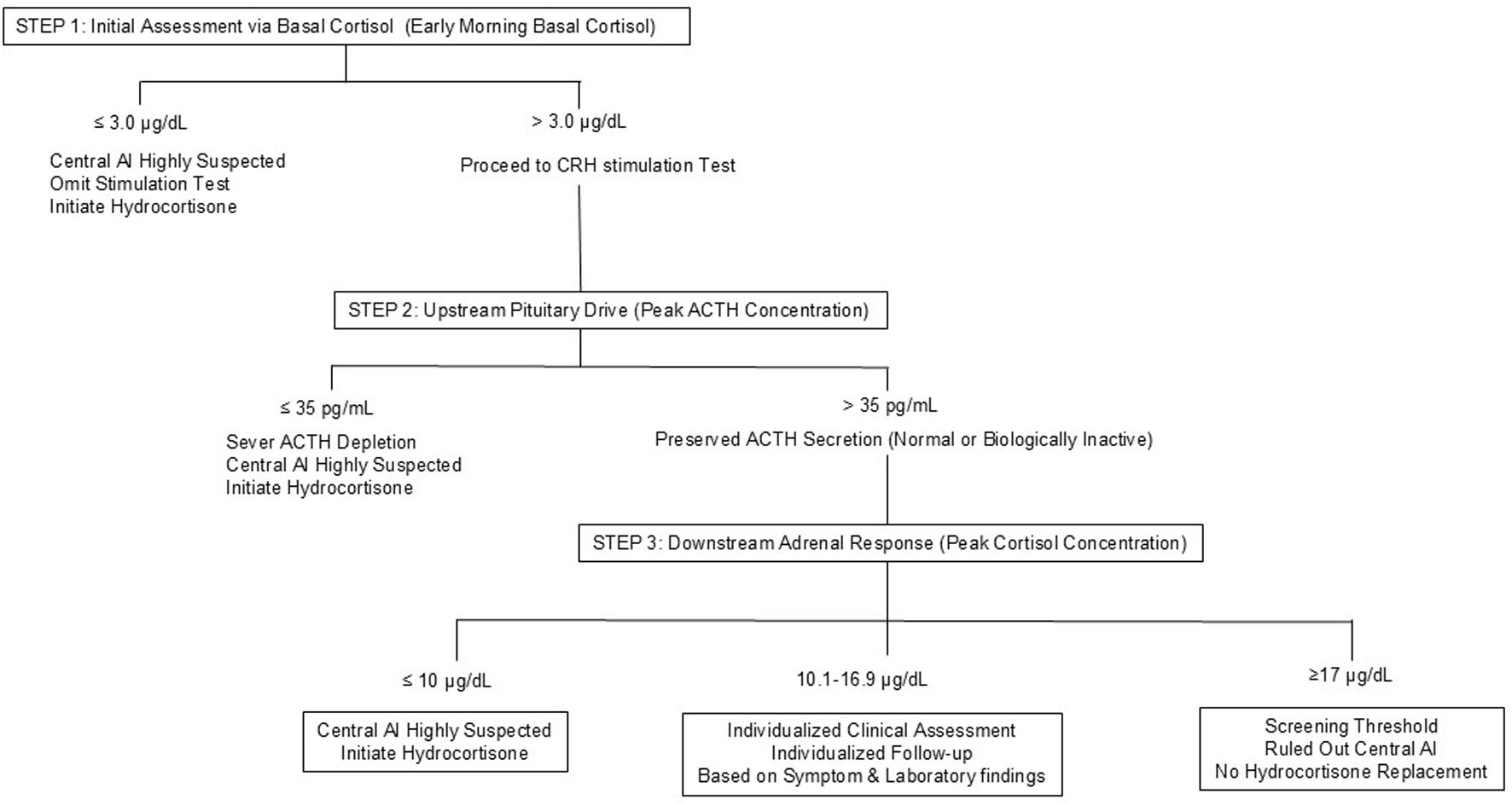 Click for large image | Figure 4. Proposed sequential clinical approach for the evaluation and management of central AI. Step 1 uses early morning basal cortisol to identify patients highly suggestive of central AI (≤ 3.0 µg/dL), for whom omission of further stimulation testing and initiation of hydrocortisone replacement may be considered. Step 2 assesses the upstream pituitary response, where a peak ACTH concentration ≤ 35 pg/mL was consistently associated with central AI in our cohort. For patients with peak ACTH concentrations > 35 pg/mL, in whom biologically inactive ACTH or exaggerated ACTH responses may be present, Step 3 evaluates the downstream adrenal response using peak cortisol concentrations. Patients are categorized into three practical groups: values ≥ 17.0 µg/dL (screening threshold), values ≤ 10.0 µg/dL (highly suggestive of central AI), and intermediate values of 10.1–16.9 µg/dL requiring individualized clinical assessment based on symptoms and laboratory findings. ACTH: adrenocorticotropic hormone; AI: adrenal insufficiency; CRH: corticotropin-releasing hormone. |
Acknowledgments
We thank the medical and co-medical staff involved in patient care. We also thank the late Dr. Kazuya Miyashita for his invaluable contributions, which greatly enriched this study, and his memory continues to inspire our work.
Financial Disclosure
This study was supported by grant 23K07982 (to K.H.) and partially funded by the Research on Rare and Intractable Disease, Health and Labour Sciences Research Grant 23FC1042 (to K.H.).
Conflict of Interest
The authors declare no conflict of interest.
Informed Consent
The requirement for written informed consent was waived because of the retrospective design, and an opt-out approach was used.
Author Contributions
M. Yoshikawa drafted the manuscript. K. Horiguchi supervised the study and revised the manuscript as the corresponding author. T. Takamizawa, S. Sekiguchi, Y. Watanuki, S. Yoshino, S. Matsumoto, R. Yamaguchi, M. Tosaka, M. Yamada, and E. Yamada contributed to data collection and provided technical support. All authors have read and agreed to the published version of the manuscript.
Data Availability
All data generated or analyzed during this study are included in this published article. Further enquiries can be directed to the corresponding author.
Abbreviations
ACTH: adrenocorticotropic hormone; AGHD: adult growth hormone deficiency; AI: adrenal insufficiency; AS: adrenal sufficiency; AUC: area under the curve; BW: body weight; CI: confidence interval; CRH: corticotropin-releasing hormone; DHEA-S: dehydroepiandrosterone sulfate; HPA: hypothalamic–pituitary–adrenal; ITT: insulin tolerance test; ROC: receiver operating characteristic; STROBE: Strengthening the Reporting of Observational Studies in Epidemiology; TRH: thyrotropin-releasing hormone
| References | ▴Top |
- Fleseriu M, Hashim IA, Karavitaki N, Melmed S, Murad MH, Salvatori R, Samuels MH. Hormonal replacement in hypopituitarism in adults: an endocrine society clinical practice guideline. J Clin Endocrinol Metab. 2016;101(11):3888-3921.
doi pubmed - Research Group on Hypothalamic-Pituitary Dysfunctions, HLSRG. Clinical practice guidelines for the diagnosis and management of hypothalamic-pituitary dysfunction, congenital arginine vasopressin resistance, and related disorders by the Japan Endocrine Society Research on Rare and Intractable Diseases. Folia Endocrinol Jpn. 2023;99:1-171. (in Japanese)
- Javorsky BR, Raff H, Carroll TB, Algeciras-Schimnich A, Singh RJ, Colon-Franco JM, Findling JW. New cutoffs for the biochemical diagnosis of adrenal insufficiency after ACTH stimulation using specific cortisol assays. J Endocr Soc. 2021;5(4):bvab022.
doi pubmed - Zha L, Li J, Krishnan SM, Brennan MR, Zhang YV, Povse P, Kerlin R, et al. New diagnostic cutoffs for adrenal insufficiency after cosyntropin stimulation using Abbott architect cortisol immunoassay. Endocr Pract. 2022;28(7):684-689.
doi pubmed - Okutan S, Jorgensen NT, Pedersen LE, Borresen SW, Hilsted L, Hansen LF, Feldt-Rasmussen U, et al. Determination of cortisol cut-off limits and steroid dynamics in the ACTH stimulation test: a comparative analysis using Roche Elecsys Cortisol II immunoassay and LC-MS/MS. Endocrine. 2024;85(1):321-330.
doi pubmed - Streeten DH, Anderson GH, Jr., Dalakos TG, Seeley D, Mallov JS, Eusebio R, Sunderlin FS, et al. Normal and abnormal function of the hypothalamic-pituitary-adrenocortical system in man. Endocr Rev. 1984;5(3):371-394.
doi pubmed - Persani L, Brabant G, Dattani M, Bonomi M, Feldt-Rasmussen U, Fliers E, Gruters A, et al. 2018 European Thyroid Association (ETA) guidelines on the diagnosis and management of central hypothyroidism. Eur Thyroid J. 2018;7(5):225-237.
doi pubmed - Izuchi T, Iwama S, Yamagami A, Kobayashi T, Suzuki K, Murase T, Ando M, et al. Defining a serum cortisol cutoff level post-CRH stimulation for diagnosing ACTH deficiency: A retrospective study validated by a nationwide registry. Front Endocrinol (Lausanne). 2026;17:1741709.
doi pubmed - Mitsui Y, Iizuka Y, Tanaka T, Hara T, Masuda S, Ohnishi Y, Kanai M, et al. An attempt to create a treatment algorithm of central adrenal insufficiency using CRH test, DHEA-S and clinical evaluation. J Med Invest. 2022;69(3.4):287-293.
doi pubmed - Allen JP, Cook DM, Olsen J. Effect of insulin induced hypoglycemia on ACTH, cortisol, and growth hormone release in man. Mil Med. 1975;140(12):837-840.
pubmed - Nelson JC, Tindall DJ, Jr. A comparison of the adrenal responses to hypoglycemia, metyrapone and ACTH. Am J Med Sci. 1978;275(2):165-172.
doi pubmed - Bailey RR, Abernethy MH, Beaven DW. Adrenocartical response to the stress of an acute myocardial infarction. Lancet. 1967;1(7497):970-973.
doi pubmed - Landon J, Wynn V, James VH. The adrenocortical response to insulin-induced hypoglycaemia. J Endocrinol. 1963;27:183-192.
doi pubmed - Arner B, Hedner P, Karlefors T, Westling H. Haemodynamic changes and adrenal function in man during induced hypoglycaemia. Acta Endocrinol (Copenh). 1963;44:430-442.
doi pubmed - Bethge H, v d Nahmer D, Zimmermann H. [The insulin hypoglycemia test as an assay of hypothalamo-hypophyseal-adrenocortical system function. I. In normal persons]. Acta Endocrinol (Copenh). 1967;54(4):668-680.
pubmed - Jasani MK, Boyle JA, Greig WR, Dalakos TG, Browning MC, Thompson A, Buchanan WW. Corticosteroid-induced suppression of the hypothalamo-pituitary-adrenal axis: observations on patients given oral corticosteroids for rheumatoid arthritis. Q J Med. 1967;36(143):261-276.
pubmed - Staub JJ, Jenkins JS, Ratcliffe JG, Landon J. Comparison of corticotrophin and corticosteroid response to lysine vasopressin, insulin, and pyrogen in man. Br Med J. 1973;1(5848):267-269.
doi pubmed - Schlaghecke R, Kornely E, Santen RT, Ridderskamp P. The effect of long-term glucocorticoid therapy on pituitary-adrenal responses to exogenous corticotropin-releasing hormone. N Engl J Med. 1992;326(4):226-230.
doi pubmed - Schmidt IL, Lahner H, Mann K, Petersenn S. Diagnosis of adrenal insufficiency: Evaluation of the corticotropin-releasing hormone test and Basal serum cortisol in comparison to the insulin tolerance test in patients with hypothalamic-pituitary-adrenal disease. J Clin Endocrinol Metab. 2003;88(9):4193-4198.
doi pubmed - Maghnie M, Uga E, Temporini F, Di Iorgi N, Secco A, Tinelli C, Papalia A, et al. Evaluation of adrenal function in patients with growth hormone deficiency and hypothalamic-pituitary disorders: comparison between insulin-induced hypoglycemia, low-dose ACTH, standard ACTH and CRH stimulation tests. Eur J Endocrinol. 2005;152(5):735-741.
doi pubmed - Dullaart RP, Pasterkamp SH, Beentjes JA, Sluiter WJ. Evaluation of adrenal function in patients with hypothalamic and pituitary disorders: comparison of serum cortisol, urinary free cortisol and the human-corticotrophin releasing hormone test with the insulin tolerance test. Clin Endocrinol (Oxf). 1999;50(4):465-471.
doi pubmed - Kazlauskaite R, Evans AT, Villabona CV, Abdu TA, Ambrosi B, Atkinson AB, Choi CH, et al. Corticotropin tests for hypothalamic-pituitary- adrenal insufficiency: a metaanalysis. J Clin Endocrinol Metab. 2008;93(11):4245-4253.
doi pubmed - de Vries F, Lobatto DJ, Bakker LEH, van Furth WR, Biermasz NR, Pereira AM. Early postoperative HPA-axis testing after pituitary tumor surgery: reliability and safety of basal cortisol and CRH test. Endocrine. 2020;67(1):161-171.
doi pubmed - Akahori H. Reversible impairment of the processing of proopiomelanocortin into ACTH in pituitary enlargement suspected of lymphocytic hypophysitis. Endocr J. 2012;59(12):1121-1129.
doi pubmed - Kobayashi A, Otsuka Y, Yoshizawa T, Tomita M, Asada H, Ikeda J, Saito M, et al. Severe hyponatremia caused by secondary adrenal insufficiency in a patient with giant pituitary prolactinoma. CEN Case Rep. 2013;2(2):184-189.
doi pubmed - Nishihara E, Kimura H, Ishimaru T, Kiriyama T, Yokoyama N, Yamashita S, Nagataki S. A case of adrenal insufficiency due to acquired hypothalamic CRH deficiency. Endocr J. 1997;44(1):121-126.
doi pubmed - Hasegawa G, Ashihara E. A case of hypothalamic adrenal insufficiency manifested normal ACTH response to insulin-induced hypoglycemia. Endocr J. 2000;47(1):51-55.
doi pubmed
This article is distributed under the terms of the Creative Commons Attribution 4.0 International License (CC BY 4.0), which permits unrestricted use, distribution, and reproduction in any medium, including commercial use, provided the original work is properly cited.
Journal of Endocrinology and Metabolism is published by Elmer Press Inc.