| Journal of Endocrinology and Metabolism, ISSN 1923-2861 print, 1923-287X online, Open Access |
| Article copyright, the authors; Journal compilation copyright, J Endocrinol Metab and Elmer Press Inc |
| Journal website https://jem.elmerpub.com |
Original Article
Volume 000, Number 000, August 2026, pages 000-000

Clinicoradiological Characteristics of Adrenal Incidentaloma: An Eight-Year Data From a Tertiary Center
Raed Aldahasha, b, c, d, Ahmed Alibrahimb, d, Meshari Al Samiha, Mohammed Alkhalafa, Nawaf Alagrafya, Yazeed Alqahtania, Abdulrahman Alqununb
aKing Saud Bin Abdulaziz University for Health Sciences, Riyadh, Saudi Arabia
bDivision of Endocrinology and Metabolism, Department of Medicine, King Abdulaziz Medical City, Riyadh, Saudi Arabia
cKing Abdullah International Medical Research Center, Riyadh, Saudi Arabia
dCorresponding Authors: Raed Aldahash, King Saud Bin Abdulaziz University for Health Sciences, Riyadh, Saudi Arabia; Ahmed Alibrahim, Division of Endocrinology and Metabolism, Department of Medicine, King Abdulaziz Medical City, Riyadh, Saudi Arabia
Manuscript submitted May 24, 2026, accepted July 21, 2026, published online August 21, 2026
Short title: Adrenal Incidentaloma
doi: https://doi.org/10.14740/jem1656
| Abstract | ▴Top |
Background: There is a rise in the incidence of adrenal incidentaloma (AI) cases owing to routine cross-sectional imaging; however, the actual practice of handling patients with AI often differs from the current international guidelines that are evidence-based. The aim of the study was to describe the clinicoradiological presentation and biochemical profile of patients with AI at a tertiary health institution in Saudi Arabia.
Methods: This retrospective cohort study involved 418 adults who had an AI of at least 1 cm in diameter for a period of 8 years. Analysis was performed regarding their clinical presentations, radiology images, hormonal assessment, and management using SPSS version 27.0. Chi-square and associated P-value were used to analyze demographics by gender.
Results: There was a predominance of females (66.5%) and high prevalence of obesity. The majority of tumors were identified incidentally (94.3%), were unilateral (92.9%), ranged between 1 to 2 cm (62.2%) in size, and exhibited a constant morphological pattern with an average follow-up period of 2.19 ± 1.56 years. On the other hand, 89.5% of baseline computed tomography studies lacked a specific protocol for the adrenal gland. Biochemically, 38.1% of patients did not pass the 1-mg overnight dexamethasone test ≥ 50 nmol/L, thus having a high incidence of mild autonomous cortisol secretion (MACS). Other types of hormonal excess were less common. Unexpectedly, only 9.3% of the total patient population was referred to the endocrinology department. Observation was chosen in 91.0% of patients, whereas 9.0% had their tumors surgically removed.
Conclusion: While most AIs were smaller and morphologically static, there were significant deficiencies in terms of guideline compliance, especially in imaging performed according to guidelines and specialist endocrinology referral. Considering that the MACS prevalence rate is quite high, it is necessary for all healthcare organizations to develop automated, multidisciplinary care pathways.
Keywords: Adrenal incidentaloma; Mild autonomous cortisol secretion; Dexamethasone suppression test; Adrenal protocol CT; Endocrinology
| Introduction | ▴Top |
Adrenal incidentalomas (AIs) are unexpected adrenal tumors usually larger than 1 cm that can be found on images taken for an imaging study that is not specifically conducted for adrenal conditions [1]. Due to the proliferation of cross-sectional imaging methods in the last decades, such as computed tomography (CT) and magnetic resonance imaging (MRI) scans, there has been a tremendous increase in the number of cases where AIs are found accidentally [2]. Despite the fact that most of these incidental adrenal mass lesions are benign non-functioning adrenocortical adenomas, the diagnosis of an AI demands a thorough systematic evaluation process [3]. Firstly, the physician needs to rule out malignancy and autonomous hormonal hypersecretion as possible causes of morbidity related to an adrenal mass lesion [4]. Malignancies comprise rare but highly fatal primary adrenocortical carcinoma, pheochromocytomas, and adrenal metastases.
In terms of autonomy, although overt symptoms associated with endocrine dysfunction are very uncommon among these patients, a considerable number of these AIs demonstrate a condition known as mild autonomous cortisol secretion (MACS) [5]. MACS has recently emerged as a condition that cannot be considered solely a benign biochemical abnormality, but as a pathophysiological state that contributes significantly to the development of serious cardiometabolic complications such as obesity, hypertension, dyslipidemia, and type 2 diabetes mellitus [6]. One important component of radiological examination involves the use of non-enhanced CT scan to ascertain the density of tissue in terms of Hounsfield Units (HU) in order to distinguish between benign and lipid-containing adenomas from a malignant and lipid-deficient adrenal mass [7]. In addition, biochemical testing should be undertaken for all patients [3].
Nevertheless, contemporary medical literature tends to emphasize the fact that there is a notable gap between protocols supported by scientific evidence and actual medical practice. Oftentimes, AIs tend to be deemed insignificant from a clinical perspective by physicians who refer the patient for further examination. These biases lead to partial imaging protocols, neglected biochemical tests, and the lack of referrals to an endocrinologist, thus putting a selected group of patients at risk of developing a cardiometabolic disease without ever being diagnosed with one.
In light of the above, this paper intends to conduct a comprehensive investigation of the clinical, imaging, and biochemical characteristics of patients presenting with AIs at a tertiary care facility over an 8-year period of time. The analysis aimed to consider follow-up dynamics, diagnostic approaches based on protocols, and rates of referrals to an endocrinologist to determine whether there are any issues regarding the treatment of AIs.
| Materials and Methods | ▴Top |
Study design and setting
The study was retrospective cohort design performed at the Ministry of National Guard Health Affairs (MNGHA) in Riyadh, Saudi Arabia. The medical records were analyzed over 8 years for the purpose of evaluating the clinical presentation, imaging features, biochemistry, and management results of patients with an AI.
Participant selection
The inclusion criteria for the participants of the study included being aged 18 years or above and having an adrenal lesion greater than or equal to 1 cm in maximum size found as an incidental finding on cross-sectional imaging studies (CT or MRI) that were not specifically ordered to investigate the presence of adrenal pathology. Exclusion criteria were active extra-adrenal malignancy, prior adrenal surgical procedure, and cross-sectional imaging ordered specifically because of overt adrenal hyperfunction. A total of 418 patients were eligible.
Data collection and variables
The data were collected by extraction from the institution’s electronic medical records system (BestCare). The variables collected were divided into three main categories: (1) Demographic and clinical data: age, gender, body mass index (BMI) defined in terms of obesity categories based on the World Health Organization classification, and type of clinical presentation, which led to the imaging (described as incidental, pressure symptom, or hyperfunctionality). (2) Radiological assessment: baseline and follow-up imaging characteristic analysis. These variables included modality of imaging (either CT or MRI), presence or absence of specific adrenal protocol (unenhanced, early, and late enhancement phases), presence or absence of lesion laterality (unilateral or bilateral), size of tumor (≤ 1, ≤ 1 to ≤ 2, ≤ 2.1 to ≤ 3, ≤ 3.1 to ≤ 4, and > 4 cm), and tissue composition (nodule/myelolipoma/lipid-rich adenoma or lipid-poor adenoma). Additionally, quantitative measurements were taken, including HU (dichotomized as < 10 HU or ≥ 10 HU) and percentage washout rate (dichotomized as < 60% or ≥ 60%). Presence or absence of an image-guided biopsy was also recorded. (3) Biochemical evaluation: The findings of biochemical tests for excessive hormone secretion were documented. The diagnosis of autonomous cortisol secretion was determined by means of 1-mg overnight dexamethasone suppression test (DST), where an absolute level of post-DST serum cortisol of 50 nmol/L (1.8 µg/dL) was taken into account to separate those with adequate suppression and autonomous secretion (≥ 50 nmol/L). Other biochemical tests were plasma adrenocorticotropic hormone (ACTH) concentrations (< 10, 10–20, > 20 pg/mL), plasma metanephrine concentrations (< 50, > 50 pg/mL), 24-h urinary metanephrines (< 2, > 2 mg/24 h), plasma renin-to-aldosterone ratio (< 2, ≥ 2), and DHEAS concentration (< 20 vs. > 20 µg/dL).
Clinical management and follow-up
The pathway of each patient was traced to determine the success in the clinical management and follow-up of cases. These comprised the formal referral of the patient for endocrine evaluation (yes or no) and the mode of management, which either involved a conservative approach such as biochemical/radiological observation and medical therapy or a surgical operation in the form of adrenalectomy. In cases where follow-up involved a longitudinal approach, the period of radiological follow-up from the initial imaging to the last one was determined.
Statistical analysis
Data analysis was conducted with the help of the IBM SPSS Statistics software package. Continuous variables, namely the length of radiological follow-up, were reported as mean ± standard deviation (SD) and range. Categorical variables were shown in numbers and percent values (n, %). The statistical comparison between two categorical variables (for instance, radiological features in different presentations or treatment strategies) was done with the use of the Chi-square test (Fisher’s exact test in case of small cell sizes). In all cases, significance was set to less than 0.05 (P < 0.05). To make sure the data were clinically and statistically valid, only complete results of laboratory and imaging studies were used for the denominator calculations per each parameter. Therefore, percentages of each sub-group were calculated taking into account the “Valid N” that denotes the total number of patients with the appropriate and available data for that particular criterion instead of the total number of patients equal to 418. The analysis was performed using SPSS version 27.0.
Ethical considerations
The protocol for this study was assessed and officially approved by the IRB of the King Abdullah International Medical Research Center (KAIMRC), Riyadh, Saudi Arabia (IRB Approval No.: 00000106525; Study Number: NRR25/039/1). This research project was given an expedited review on January 23, 2025. The study was conducted in compliance with the ethical standards of the responsible institution for human subjects and in accordance with the Declaration of Helsinki. The researcher team fully abided by the Saudi Law of Ethics of Research on Living Creatures and their regulations.
| Results | ▴Top |
Demographics and baseline characteristics
The total number of participants included in the study was 418. The demographic characteristics of the cohort can be seen in Table 1. In terms of gender, females were the majority (66.5%, n = 278) compared to men (33.5%, n = 140). Age at presentation showed that the disease was more common in the older population. Those above 60 years old were the most numerous with a proportion of 37.0% (n = 74), followed by those who were between 51 and 60 years old (29.5%, n = 59) and those who were between 41 and 50 years old (25.5%, n = 51). The occurrence among the younger ones below 40 years old was very rare with a percentage of just 8.0% of those whose ages were available. The BMI values were obtained for 195 patients, and 53.3% (n = 223) did not have BMI data. Compared to the whole population in the study, some of the participants exhibited high BMI readings where 29.1% (n = 122) were obese (covering obesity classes I, II, and III), and 11.7% (n = 49) were overweight. Those with normal BMI were very few (5.5%, n = 23) and there was even one participant (0.2%) who was underweight (Table 1).
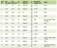 Click to view | Table 1. Demographic Characteristics of the Subjects (Categorical) |
Distribution of demographic characteristics by gender
According to Table 2, analysis through cross-tabulation showed that there were different characteristics regarding age and BMI among patients based on their gender. There was no significant difference in the distribution of age among the patients regardless of whether they were female or male (P = 0.253). The majority of the participants (70.5% females and 68.6% males) were aged 60 years or older. On the other hand, there was a significant difference in the distribution of the BMI among the patients irrespective of whether they were female or male (P = 0.040). More male patients than female patients were overweight (16.4% vs. 9.4%).
Click to view | Table 2. Distribution of Demographic Characteristics by Gender |
Baseline clinical, radiological, and biochemical characteristics
The initial clinical presentation leading to imaging was identifiable for 176 patients (42.1% of the total cohort). Among this subgroup with available data, the majority of adrenal masses were incidentally found (94.3%, n = 166), while a small proportion presented with hyperfunctionality (3.4%, n = 6) or pressure symptoms (2.3%, n = 4) (Table 3). CT scanning emerged as the main technique used for the diagnosis (96.7%, n = 380); nevertheless, the use of an adrenal-specific CT protocol was applied only in 10.5% (n = 41) of cases. The majority of radiographic findings corresponded to solitary adrenal lesions (92.9%, n = 169); furthermore, most lesions were relatively small, with a size between 1 and 2 cm (62.2%, n = 112). Large lesions larger than 4 cm were uncommon (4.4%, n = 8). In terms of radiological appearance, most lesions were characterized by being rich in fat (96.7%, n = 175). In agreement with these radiological findings, low attenuation of the lesion was seen on unenhanced CT scans in 63.9% (n = 78) of cases, whereas absolute washout lower than 60% was identified in 83.2% (n = 79). Because of the presence of distinctive imaging features, few invasive diagnostic tests had to be done, specifically biopsies, which were carried out in 6.3% (n = 9) of cases (Table 3).
Click to view | Table 3. Baseline Clinical, Radiological, and Biochemical Characteristics of the Subjects |
The biochemical analysis indicated that most of the patients did not have a problem with hormonal hypersecretion. After administering a 1 mg DST, about 61.9% (n = 91) of the tested patients were found to have a suppressed value of cortisol (< 50), but there was a considerable number who had ≥ 50 levels of ACTH levels. ACTH levels were found relatively evenly spread, with 36.2% (n = 34) showing ACTH < 10, 27.7% (n = 26) showing an ACTH level of 10–20, and 36.2% (n = 34) having ACTH > 20. Pheochromocytoma was rare, with only plasma metanephrines being less than 50 in 84.5% (n = 87) and urinary metanephrines < 2 in 95.2% (n = 59). Primary aldosteronism and androgen excess were also rare, as illustrated in Table 3.
Follow-up radiological characteristics
Table 4 displays the features of the radiological follow-up findings. The average time of radiological follow-up among this cohort was 2.19 ± 1.56 years. Throughout the period of radiological follow-up, CT was used in 96.0% (n = 97) of cases as the primary radiological modality, while only 4.0% (n = 4) used MRI for imaging. Similar to baseline examination, most follow-up examinations using CT did not use the specific adrenal protocol, which is applied in 92.8% (n = 90) of cases. From the radiological follow-up evaluation, the adrenal lesions were found to remain predominantly unilateral, comprising 92.2% (n = 59) of cases. In most cases, the adrenal adenoma size was 1–3 cm, with 50.0% (n = 38) of adenomas being 1–2 cm and 32.9% (n = 25) ranging from 2.1 to 3 cm. Lesions that exceeded 4 cm in diameter accounted for 2.6% (n = 2) of cases. Regarding tissue composition, 95.7% (n = 67) of the masses maintained imaging characteristics consistent with lipid-rich adenomas, nodules, or myelolipomas. Unenhanced CT attenuation evaluations revealed a relatively even split, with 54.5% (n = 18) of lesions presenting with densities ≥ 10 HU and 45.5% (n = 15) measuring < 10 HU. Among the subset of patients evaluated for contrast washout, 71.4% (n = 20) demonstrated an absolute washout of < 60%, while 28.6% (n = 8) exhibited a washout of ≥ 60%.
Click to view | Table 4. Follow-Up Radiological Characteristics of the Subjects |
Referral patterns and clinical management
Assessment of the use of clinical pathways indicated that nearly all of the patients diagnosed with AI had been treated without any consultation from an endocrinologist. As shown in Table 5, of the entire pool of patients (N = 418), only 9.3% (n = 39) had been referred to an endocrinologist, while the rest (90.7%) (n = 379) had not been referred. In terms of treatment options, information was available for 199 patients. Among those, management through a conservative approach dominated, accounting for 91.0% (n = 181). Surgery was considered for only a few patients (9.0%) (n = 18).
Click to view | Table 5. Referral and Management of the Subjects |
| Discussion | ▴Top |
Key observations from this 8-year retrospective study from a tertiary center have revealed important trends in the clinical, radiological, and biochemical profile of AI, along with a number of important lacunae in their standardized management. The patient population of 418 included in this study consisted mostly of elderly individuals, with the overwhelming majority above the age of 50 years, consistent with previous studies. In addition, a large number of the patients belonged to the female gender and had overweight or obese BMI with females having significantly greater class III obesity compared to males. From the radiological aspect, a majority of the patients with AIs showed a predominance of small-size (1–2 cm), unilateral, and lipid-rich adrenal masses that remained constant during the follow-up of 2.19 years. Nevertheless, we found several deficiencies in the proper use of diagnostic guidelines for AI, as evidenced by almost 90% of the initial CT scans not following the adrenal protocol and merely 9.3% being referred to endocrinologists for further investigations. Notably, despite the exceptionally low referral rate, approximately 40% of the patients who underwent DST had MACS.
The female preponderance (66.5%) and elderly age group found in this study have excellent agreement with current global evidence. An increased incidence rate of AIs with increasing age is known in literature and can be accounted for due to either genetic mutations related to aging in adrenal glands or increased cross-sectional imaging in elderly population [8]. It is interesting to mention that there were a considerable number of obese patients identified in our patient population; 30% of our cases had obesity based on the available information. The association between adrenocortical neoplasms and obesity is not only based on the increased possibility of performing imaging tests in obese patients. According to current scientific research, a bidirectional relationship exists between metabolic disorders and adrenal enlargement. Adenomas and AIs are linked to insulin resistance [9].
In terms of mechanistic pathways, insulin resistance will result in compensatory hyperinsulinemia, with mitogenic effects in the adrenal cortex through activation of the insulin-like growth factor system. Both insulin and insulin-like growth factor 1 (IGF-1) receptors are abundantly found in adrenal cortical cells. Under conditions of obesity, IGF-1 is increased in terms of bioavailability because of changes in the production of IGF-binding proteins in the liver, and therefore, cell proliferation while inhibiting apoptosis will occur within the adrenal gland [10]. Additionally, the presence of visceral fat will create a pro-inflammatory environment by having increased adipokines such as interleukin-6 and tumor necrosis factor-alpha, causing stimulation of hyperplasia in the adrenal cortex. Hence, the prevalence of obesity within our study population might not just be a concomitant condition, but a key determinant in AI pathology.
In spite of the noted morphological homogeneity of the tumors found in our patients, the radiologic evaluation initially performed was suboptimal. According to ESE/ENSAT guidelines, the assessment of HU density using an unenhanced CT scan is the primary diagnostic approach for excluding malignancy [11]. Yet, 89.5% of baseline and 92.8% of follow-up scans did not comply with an adrenal protocol, leading to a major loss of critical HU and washout values. This consistent failure at a tertiary center likely stems from several operational factors. First, a knowledge gap may exist among non-endocrine ordering physicians who may classify AIs as “clinically insignificant” findings rather than tumors requiring specific imaging phenotypes. Second, the electronic medical record (EMR) system (BestCare) currently lacks automated prompts or a dedicated “adrenal protocol” order set that triggers when an adrenal mass is identified by a radiologist. Finally, there may be a lack of standardized radiologist reporting where the discovery of a mass does not explicitly prompt a recommendation for a protocol-driven re-evaluation. Without this structured approach, the risk of missing lipid-poor adenomas or potential malignant progression remains high, directly contributing to the low endocrinology referral rate (9.3%) and the systemic underdiagnosis of hormonal hypersecretion observed in this cohort.
One of the most troubling findings related to the endocrinology referral rate is the lack of proper identification of MACS. Among patients who had undergone 1 mg overnight DST in our study population, an alarming 38.1% failed to suppress their cortisol below 50 nmol/L (1.8 µg/dL). MACS is no longer considered a harmless or “subclinical” entity but rather a disease characterized by chronic and low-level hypercortisolism [12]. Several recent studies involving a high number of participants clearly showed that MACS has a dramatically higher cardiometabolic risk profile, and people suffering from MACS have higher rates of resistant hypertension, type 2 diabetes mellitus, dyslipidemia, and osteoporotic bone fractures than patients with purely non-functioning tumors [13–15]. Thus, by not referring these patients to specialized endocrinologic teams, a considerable portion of our population may have been deprived of important treatments. Proper identification of MACS is crucial, although not necessarily surgical removal of the tumor, which was conducted only in 9.0% of our sample group. Instead, MACS should serve as an indicator for the identification and medical treatment of silent cardiometabolic disorders.
Strengths and limitations
Some of the strengths of the study include the considerable sample size as well as the relatively long observational period of 8 years that give an accurate picture of the real-life scenario concerning AI treatment and care at one of the most important centers in Saudi Arabia. Nevertheless, the study has some weaknesses as well. First and foremost, there was a considerable amount of missing data because of the nature of the retrospective study design. This was particularly evident regarding initial clinical presentation (missing in 57.9% of cases), BMI measurements, HU quantification, and biochemical tests, which limits the ability to generalize these specific clinical characteristics to the entire cohort. Secondly, being a tertiary center, the study might have been biased by referrals, but strangely enough, the number of internal referrals for endocrinology patients was rather small. Thirdly, the results do not have any longitudinal data to prove the relationship between MACS and mortality.
Conclusion and Implications
Despite the predominance of small, benign, and stable adrenals among patients with AI in this study group, it was noted that the clinical approach involved severe insufficiency regarding protocol-based radiological imaging and biochemical workups. Considering the significant prevalence of obesity and possible autonomous cortisol secretion (MACS) among patients with AI, there is evidence of the systemic metabolic risks of having AIs. This information has practical applications in clinical practice. It is essential to develop multidisciplinary institutional protocols and pathways that automatically trigger endocrinology consultations with required imaging protocols in every patient with incidentally found adrenal mass. This gap should be filled to ensure early identification of MACS and optimization of management of associated cardiometabolic disorders, as well as avoid unnecessary financial and mental burden due to radiological surveillance of benign adrenal tumors.
Acknowledgments
None to declare.
Financial Disclosure
None to declare.
Conflict of Interest
None to declare.
Informed Consent
Because this research used de-identified electronic health records, the IRB exempted this research project from obtaining the participant’s written consent.
Author Contributions
Raed Aldahash and Ahmed Alibrahim conceptualized and supervised the study. Nawaf Alagrafy, Meshari Al Samih, and Mohammed Alkhalaf contributed to data collection and methodology development. Yazeed Alqahtani and Abdulrahman Alqunun performed data analysis and literature review. All authors contributed to manuscript drafting, critical revision, and approved the final version of the manuscript. Raed Aldahash and Ahmed Alibrahim served as corresponding authors and were responsible for project administration and communication with the journal
Data Availability
The authors declare that data supporting the findings of this study are available within the article.
| References | ▴Top |
- Sconfienza E, Tetti M, Forestiero V, Veglio F, Mulatero P, Monticone S. Prevalence of functioning adrenal incidentalomas: a systematic review and meta-analysis. J Clin Endocrinol Metab. 2023;108(7):1813-1823.
doi pubmed - Cuthbertson DJ, Alam U, Davison AS, Belfield J, Shore SL, Vinjamuri S. Investigation and assessment of adrenal incidentalomas. Clin Med (Lond). 2023;23(2):135-140.
doi pubmed - Owei L, Wachtel H. The landmark series: evaluation and management of adrenal incidentalomas. Ann Surg Oncol. 2025;32(7):4712-4719.
doi pubmed - Chatzellis E, Kaltsas G. Adrenal Incidentaloma. In: Feingold KR, Adler RA, Ahmed SF, Anawalt B, Blackman MR, Chrousos G, et al., eds. Endotext. South Dartmouth (MA). 2000.
pubmed - Turan Erdogan B, Evranos Ogmen B, Sacikara M, Aydin C, Topaloglu O, Ersoy R, Cakir B. The relationship between mild autonomous cortisol secretion and metabolic diseases in cases with adrenal incidentaloma. Endokrynol Pol. 2025;76(2):172-181.
doi pubmed - Prinzi A, Lombardo AM, Finocchiaro S, Galvano A, Vella V, Frasca F, Malandrino P. Expanding the clinical profile of mild autonomous cortisol secretion: new diagnostic markers and emerging complications. Endocr Pract. 2026;32(1):46-52.
doi pubmed - Gasmi A, Taieb A, Hattab F, Ghachem A, Ben Haj Slama N, Halloul I, Saafi W, et al. Characterization of unilateral adrenal incidentalomas: hormonal analysis, computed tomography, and magnetic resonance imaging correlation. Cureus. 2025;17(12):e100542.
doi pubmed - Kebebew E. Adrenal Incidentaloma. N Engl J Med. 2021;384(16):1542-1551.
doi pubmed - Terzolo M, Pia A, Ali A, Osella G, Reimondo G, Bovio S, Daffara F, et al. Adrenal incidentaloma: a new cause of the metabolic syndrome? J Clin Endocrinol Metab. 2002;87(3):998-1003.
doi pubmed - Lazurova I, Jochmanova I, Sotak S, Spakova I, Marekova M. Is there a role for the IGF system and epidermal growth factor (EGF) in the pathogenesis of adrenocortical adenomas? A preliminary case-control study. Physiol Res. 2020;69(6):1085-1094.
doi pubmed - Fassnacht M, Tsagarakis S, Terzolo M, Tabarin A, Sahdev A, Newell-Price J, Pelsma I, et al. European Society of Endocrinology clinical practice guidelines on the management of adrenal incidentalomas, in collaboration with the European Network for the Study of Adrenal Tumors. Eur J Endocrinol. 2023;189(1):G1-G42.
doi pubmed - Prete A, Bancos I. Mild autonomous cortisol secretion: pathophysiology, comorbidities and management approaches. Nat Rev Endocrinol. 2024;20(8):460-473.
doi pubmed - Iriarte-Duran MB, Donato S, Herrera A, Vega A, Jimenez Casinello JM, Marazuela M, Araujo-Castro M. The impact of mild autonomous cortisol secretion and proposed interventions. Expert Rev Endocrinol Metab. 2025;20(4):251-266.
doi pubmed - Czapla-Iskrzycka A, Swiatkowska-Stodulska R, Sworczak K. Comorbidities in mild autonomous cortisol secretion - a clinical review of literature. Exp Clin Endocrinol Diabetes. 2022;130(9):567-576.
doi pubmed - Jimenez Cassinello JM, Vega-Beyhart A, Bernarda Iriarte M, Donato S, Herrera-Martinez AD, Marazuela M, Araujo-Castro M. Mild autonomous cortisol secretion: impact on bone health and quality of life. A review. Endocrine. 2025;88(3):693-700.
doi pubmed
This article is distributed under the terms of the Creative Commons Attribution 4.0 International License (CC BY 4.0), which permits unrestricted use, distribution, and reproduction in any medium, including commercial use, provided the original work is properly cited.
Journal of Endocrinology and Metabolism is published by Elmer Press Inc.