| Journal of Endocrinology and Metabolism, ISSN 1923-2861 print, 1923-287X online, Open Access |
| Article copyright, the authors; Journal compilation copyright, J Endocrinol Metab and Elmer Press Inc |
| Journal website https://jem.elmerpub.com |
Original Article
Volume 16, Number 5, October 2026, pages 228-237

Evaluation of Hypothalamic–Pituitary–Adrenal Axis After Short Course of Systemic Steroids
Pooja Jadhaoa, Sravya Surapanenia, Swayamsidha Mangaraja, Abhay Sahooa, Jayshree Swaina, Subhashree Rayb, Bikash Ranjan Karc, Jaya Bhanu Kanwara, d
aDepartment of Endocrinology, IMS and SUM Hospital, Bhubaneswar, Odisha, India
bDepartment of Biochemistry, IMS and SUM Hospital, Bhubaneswar, Odisha, India
cDepartment of Dermatology, IMS and SUM Hospital, Bhubaneswar, Odisha, India
dCorresponding Author: Jaya Bhanu Kanwar, Department of Endocrinology, IMS and SUM Hospital, Bhubaneswar, Odisha, India
Manuscript submitted July 23, 2026, accepted September 16, 2026, published online October 31, 2026
Short title: HPA Axis After Systemic Steroid Use
doi: https://doi.org/10.14740/jem1662
| Abstract | ▴Top |
Background: Hypothalamic–pituitary–adrenal (HPA) axis suppression is considered an adverse effect of long-term glucocorticoid treatment. However, short-term steroid therapy may also suppress the HPA axis. The objective of this study is to examine the temporal pattern of HPA axis suppression and recovery after a short duration of glucocorticoids and recognize predictors of HPA axis suppression.
Methods: A total of 229 patients visiting outpatient departments at a tertiary care center and being prescribed steroids for various underlying conditions were recruited, of whom 121 patients completed the study. Testing of the HPA axis was done by an 8 am serum cortisol concentration and 1-µg adrenocorticotrophic hormone (ACTH)-stimulated cortisol (when needed), measured at baseline before starting steroid therapy and after completion of therapy on days 3, 10, 17, 24, and 31.
Results: HPA axis suppression was seen in 57 (47%) study subjects on day 3 post-steroid course. On day 10, 18 (31.5%) subjects had persistent suppression, while 39 (68.5%) subjects recovered. All patients had recovery of the HPA axis by day 31. Logistic regression analysis revealed lower odds of HPA axis suppression on day 3 after completion of steroid therapy with age of the patient (P = 0.018) and baseline cortisol before starting steroid therapy (P = 0.036). Glucocorticoid tapering showed a protective direction of association but did not reach statistical significance.
Conclusion: HPA axis suppression after a short course of steroid therapy is transient with patients recovering within 1 month after completion of steroid therapy. Younger age and lower baseline morning cortisol were independently associated with a greater likelihood of HPA axis suppression.
Keywords: HPA axis suppression; Short-term steroid therapy; Adrenal insufficiency; Glucocorticoid treatment; Recovery predictors
| Introduction | ▴Top |
The most frequent cause of adrenal suppression is the use of exogenous corticosteroids. Glucocorticoid-induced adrenal insufficiency occurs as a result of suppression of the hypothalamic–pituitary–adrenal (HPA) axis leading to adrenal insufficiency. The HPA axis may remain suppressed after discontinuation of steroid therapy, leaving the patient vulnerable to an adrenal crisis [1]. There appears to be a discrepancy between the high prevalence of biochemically detected glucocorticoid-induced adrenal insufficiency and the relatively low reporting of its clinical manifestations, indicating a possible under-recognition of the condition [2]. A systematic review and meta-analysis of 73 studies reported a median prevalence of glucocorticoid-induced adrenal insufficiency of 37% among patients receiving glucocorticoid therapy in any form [3]. Another meta-analysis found median prevalence rates of 48.7% in patients treated with oral glucocorticoids and 52.2% in those receiving intra-articular injections [4]. Identification of HPA axis suppression leading to adrenal insufficiency is essential, as it can result in a life-threatening adrenal crisis, particularly during periods of stress or underlying infection [5]. It is a severe complication of adrenal insufficiency with 15.1 per 100 patient-years as the incidence rate in patients with glucocorticoid-induced adrenal insufficiency and mortality of almost 6% per adrenal crisis events [6, 7]. Hence, it is imperative to take cognizance of HPA axis suppression and prevent it. Given the widespread use of systemic steroids and the substantial individual variation in susceptibility to glucocorticoid-induced adrenal insufficiency, it is crucial to identify valid predictors of diagnosis, recovery, and clinical adverse outcomes. The identified predictors of adrenal insufficiency could potentially stratify patients into risk categories and enable appropriate management [8]. However, there is no absolute cut-off value for the dose of steroid taken, duration of treatment, route of administration or type of steroid taken that predict adrenal suppression [9]. There is major variability in the inter-individual response to steroids probably related to different genetic profiles which regulate glucocorticoid receptor activity [10]. Adrenal insufficiency is a frequent consequence of prolonged glucocorticoid therapy [11, 12]. Although short-term (less than 2 weeks) high-dose glucocorticoid use is generally associated with rapid recovery of adrenal function [13, 14], several studies have reported suppression of the HPA axis even after brief courses of glucocorticoid treatment [15–17]. In a prospective study involving 311 patients with chronic obstructive pulmonary disease (COPD) exacerbations treated with 40 mg of prednisolone for ≤14 days, abrupt discontinuation of the drug was found to be safe [16]. In a prospective study of cancer patients who received at least three short courses of high-dose dexamethasone (3–4 days each) every 2–4 weeks, 15% developed glucocorticoid-induced adrenal insufficiency within 3–6 months [17]. A systematic review of 10 studies involving children with acute lymphoblastic leukemia found that glucocorticoid-induced adrenal insufficiency occurred in nearly all patients treated with high-dose systemic glucocorticoids for less than 2 months. Most children regained adrenal function within a few weeks; however, the exact duration of recovery remains uncertain [18]. General consensus agrees to anticipate adrenal insufficiency in patients taking > 7.5 mg/day of prednisolone for 3 weeks and it is suggested that steroids can be withdrawn if treatment duration is less than 3 weeks [19]. Therefore, most practicing clinicians will withdraw steroids after short-term therapy without tapering. Despite the risks associated with HPA axis suppression, not many studies have evaluated the exact temporal pattern of HPA axis suppression and recovery after a short course of glucocorticoids. Our study aimed to evaluate prospectively the status of HPA axis after a short course of systemic steroid treatment.
| Materials and Methods | ▴Top |
We conducted a prospective cohort study in a large tertiary care teaching hospital with the objective to evaluate the time to suppression and the duration of HPA axis suppression after a short course of systemic steroid treatment and to define predictors of HPA axis suppression and recovery (which includes the duration of steroid therapy, cumulative dose of steroid, the type of steroid used or the method of cessation of steroids—tapered or not). The study was conducted for 18 months from November 2021 to April 2023.
We included all patients who were prescribed steroids for a short duration between 5 and 45 days for various underlying conditions such as respiratory (COPD, asthma), neurological (cerebral edema, Bell’s palsy, paraparesis), dermatological (acute dermatitis, eczema, anaphylaxis), hematological (malignancy, auto-immune hemolytic anemia (AIHA)) or rheumatological disorders (vasculitis) and receiving more than the replacement dose (> 7.5 mg of prednisolone equivalents/day). We excluded all patients with a history of previous glucocorticoid intake within the last 6 months, inhalational steroid intake within the last 3 months, Addison’s disease, adrenal metastasis, pituitary adenoma/Cushing’s disease, hypothalamic or pituitary surgery, central nervous system (CNS) tumor irradiation, critical illness-related adrenal insufficiency, past history of adrenal tuberculosis, disseminated fungal infection, drug intake—ketoconazole, imipramine, chlorpromazine, and non-consenting individuals. This study was performed in line with the principles of the Declaration of Helsinki. Ethical approval was obtained from the Institutional Ethics Committee of Siksha O Anusandhan University (DMR/IMS.SH/SOA/2021/121) and a written informed consent was obtained from all participants before enrolment and study procedures.
Serum cortisol (8 am), serum sodium, potassium, serum urea, creatinine, and blood glucose were measured at baseline before starting steroid therapy. Serum cortisol was measured by the electrochemiluminescence immunoassay (ECLIA) in cobas e411 modular analyzer (Roche Diagnostics GmbH, Sandhofer Strasse 116, D-68305 Mannheim) with a range of assay of 0.054–63.4 µg/dL and coefficient of variance of 4.5%. To assess adrenal insufficiency, we assessed “baseline cortisol” which was fasting 8 am cortisol concentration. After an overnight 8-h fast, when the morning 8 am sample of serum cortisol was less than 3.5 µg/dL, the HPA axis was said to be suppressed according to guidelines by the Endocrine Society [5]. If the morning serum cortisol concentration was more than 12.5 µg/dL, then the axis was said to be not suppressed [20]. If the morning serum cortisol concentration was between 3.5 and 12.5 µg/dL, then the 1-µg adrenocorticotrophic hormone (ACTH) stimulation test (low-dose short synacthen test) was done. In their study, Sagar et al concluded that all patients who underwent the stimulation test, who had 8 am cortisol concentration of more than 12.5 µg/dL, passed the stimulation test. A 1-µg dose of ACTH was prepared from a 250-µg vial of Syntropac (tetracosactide acetate, ACTH 1–24) by diluting it in 100 mL of 0.9% normal saline. Of the prepared solution, 0.4 mL (i.e., 16 units in a 40-IU insulin syringe) which has a concentration of 1 µg/mL of synacthen was given intravenously and serum cortisol concentration was tested after 30 min [21, 22]. This sample was labeled as “stimulated cortisol”, and if the post-stimulation cortisol concentration was less than 18.5 µg/dL, the response was considered indicative of HPA axis suppression. Patients were evaluated on the third day after completing glucocorticoid therapy, followed by weekly assessments for 4 consecutive weeks to monitor recovery of the HPA axis. No imputation of missing biochemical observations was performed. Participants who did not complete the predefined follow-up protocol were excluded from the final analysis. The study plan is illustrated in Figure 1.
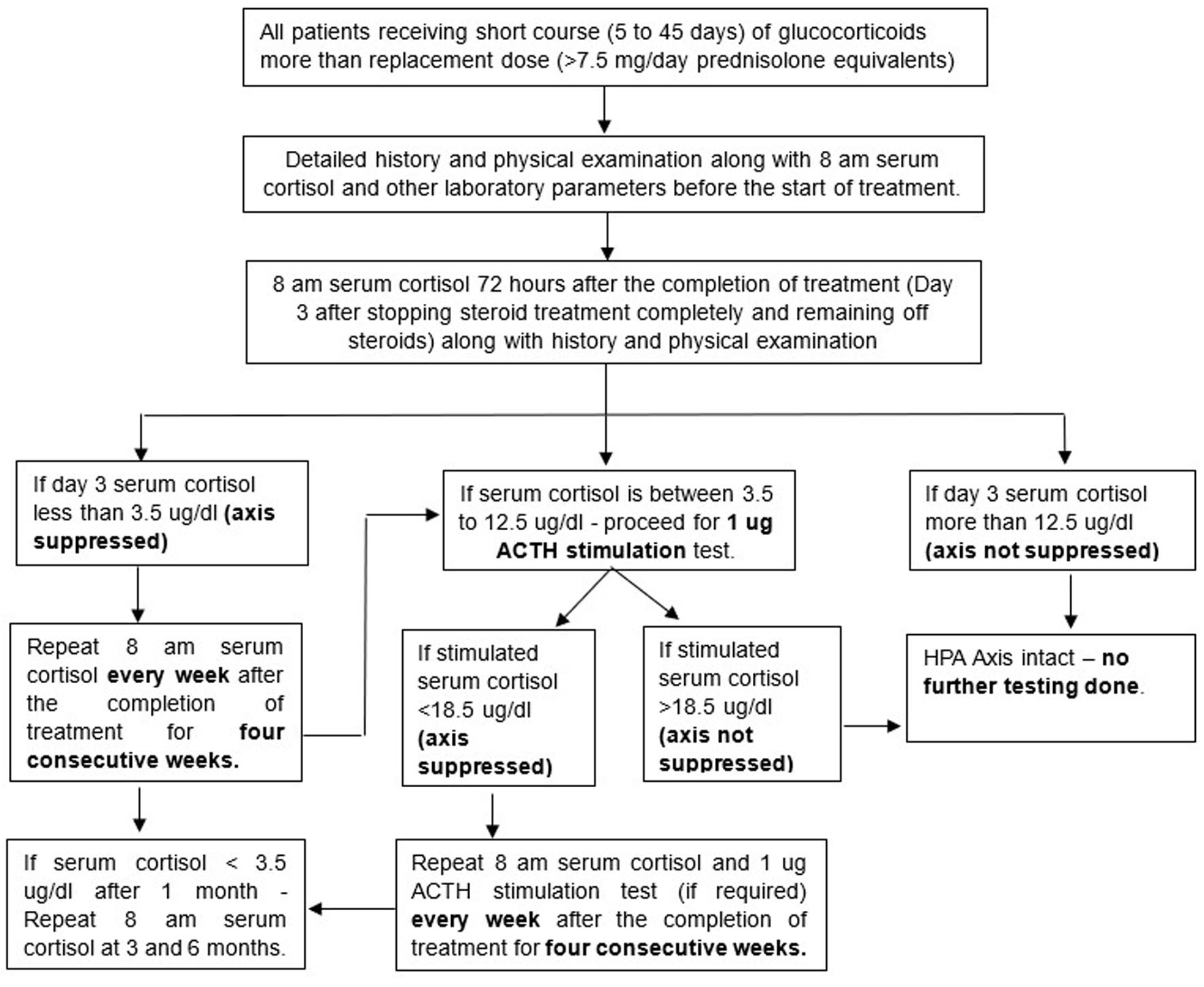 Click for large image | Figure 1. Flow chart of the study plan. |
Statistical analysis
Based on the findings of a previous study conducted by Henzen et al. [15], if we consider the proportion of patients with suppressed HPA axis as 45.3%, then a sample size of 96 subjects is needed to estimate the true value with 10% error and 95% confidence interval (CI). Considering a dropout rate of 10%, our sample size was calculated to be 106 patients.
Data were analyzed using the IBM SPSS statistical software package, version 29. Normality of the data was checked using the Kolmogorov-Smirnov test. Student’s t-test or Mann-Whitney U-test was used where appropriate and for categorical variables the Chi-squared test was used. Logistic regression analysis was done to determine predictors of HPA axis suppression. The statistical significance was measured at the 5% level of significance and P value > 0.05 was considered statistically significant.
| Results | ▴Top |
A total of 229 subjects were started on short-term steroids in various outpatient departments of our hospital of whom 121 subjects completed the study and were included in the analysis, as shown in Figure 2. Forty percent of the subjects were from the Department of Dermatology, while 29% were from the Department of Neurology. The Departments of Pulmonary Medicine and Otolaryngology contributed 10% and 6.6% of the subjects, respectively. The Departments of Ophthalmology, Orthopedics, and Hematology each accounted for approximately 4% of the study population. The mean age of our study group was 45.07 ± 14.61 years of whom 53% were females and 47% were males. The mean body mass index (BMI) of our study subjects was 24.95 ± 3.44 kg/m2 and mean baseline serum cortisol concentration of patients before starting the steroid treatment was 11.12 ± 2.91 µg/dL. The mean cumulative dose of steroids of all study subjects was 858.6 ± 1,334.64 mg, with cumulative exposure ranging from 100 to 6,250 mg prednisolone equivalent dose and the mean total number of days for which steroids were taken was 15.98 ± 9.51 days (ranging from 5 to 45 days). Maximum 40% of subjects consumed total steroid dose between 251 and 500 mg prednisolone equivalent dose. Cumulative steroid doses equivalent to 100–250 mg, 501–1,000 mg, and more than 1,000 mg of prednisolone were observed in 25%, 20%, and 15% of the subjects, respectively. Study subjects with a total duration of steroid consumption between 5 and 10 days, 11 and 15 days, 16 and 20 days and more than 20 days were 47%, 16%, 17%, and 20%. The most commonly used steroid preparation was prednisolone, used in 46% of total study subjects, followed by methylprednisolone, used in 33% of study subjects. All the study subjects (121 patients) were tested on day 3 after stopping the steroid course. Biochemical HPA axis suppression occurred in 57 of 121 participants (47.1%) on day 3. The corresponding 95% CI is approximately 38.4–56.0%.
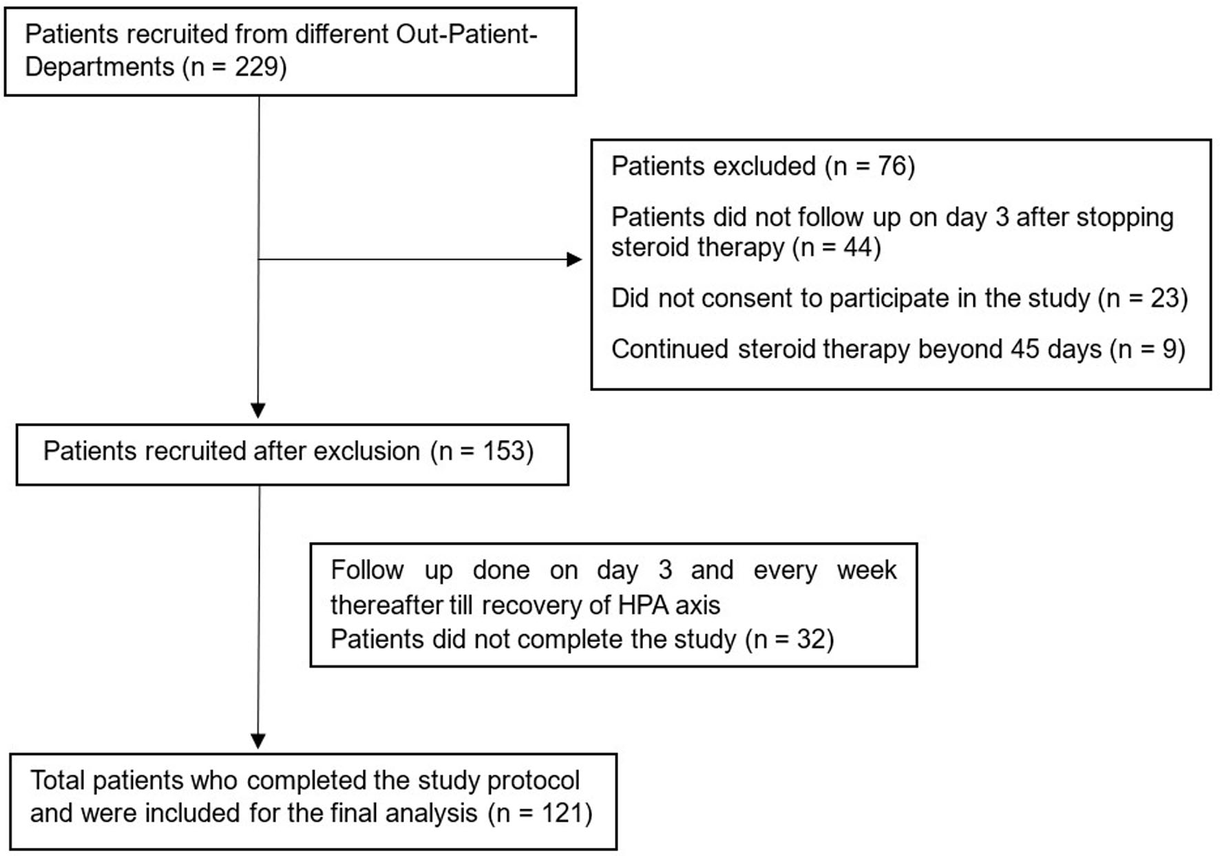 Click for large image | Figure 2. Study subject distribution. |
All 121 study participants initially underwent assessment with 8 am serum cortisol, and the 1-µg ACTH stimulation test was performed only in those with intermediate cortisol concentration (3.5–12.5 µg/dL). On day 3 after completion of steroid therapy, 43 of the 57 patients classified as having HPA axis suppression were identified based on the suppressed 8 am cortisol concentration, whereas 14 additional patients were classified as having suppressed HPA axis based on an abnormal response to the 1-µg ACTH stimulation test because of having intermediate basal cortisol concentration. Patients with morning cortisol concentration > 12.5 µg/dL were considered to have an intact HPA axis and did not undergo stimulation testing. Thus, of the 57 patients, 43 patients had suppressed 8 am cortisol and 14 patients had suppressed 1-µg ACTH stimulated cortisol. A total of 57 subjects (who had suppressed HPA axis on day 3 after completion of steroid course) were retested one week later (that is on day 10 after stopping the steroid). Out of the 57 subjects, 18 subjects had persistent HPA axis suppression and 39 subjects had recovery of the HPA axis. A total of 18 subjects (who had suppressed HPA axis on day 10 after completion of the steroid course) were retested one week later (that is on day 17 after stopping the steroid). Out of the 18 subjects, four subjects had persistent HPA axis suppression and 14 subjects had recovery of the HPA axis. The remaining four subjects were retested one week later (on day 24 after stopping the steroid). Among these four patients, three patients had recovery of the HPA axis and one patient with persistent HPA axis suppression was retested 1 week later on day 31 after completion of the steroid course and was found to have recovery of the HPA axis, as shown in Table 1.
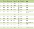 Click to view | Table 1. Status of HPA Axis During the Study Period |
At each follow-up visit, participants underwent clinical assessment for symptoms suggestive of adrenal insufficiency. Given that clinical manifestations of adrenal insufficiency are uncommon after a short course of glucocorticoid therapy, our study focused on assessing biochemical HPA axis suppression using basal morning cortisol measurements and the 1-µg ACTH stimulation test. Although biochemical HPA axis suppression was identified in 57 patients, none developed adrenal crisis or clinically overt adrenal insufficiency requiring hospitalization or emergency glucocorticoid therapy. Symptoms such as fatigue and generalized weakness were occasionally reported but were difficult to distinguish from manifestations of the underlying primary disease and therefore were not analyzed as study outcomes.
Study subjects with HPA axis suppression on day 3 were significantly younger than those who did not have HPA axis suppression and had significantly lower baseline (before starting the steroid therapy) 8 am serum cortisol concentration. It was seen that the cumulative dose of steroid was significantly lower (550.18 ± 896.47 mg vs. 1,133.28 ± 1,586.12 mg) in the subjects with HPA axis suppression as compared to those without HPA axis suppression, as shown in Table 2. HPA axis suppression was significantly lower in subjects where steroids were tapered before discontinuation (Table 2). Overall, 61 of 121 participants (50.4%) underwent tapering of corticosteroid therapy before discontinuation. Tapering was performed in 21/57 (36.8%) participants with HPA axis suppression and 40/64 (62.5%) participants without HPA axis suppression (P = 0.006). There was no uniform tapering regimen across the study population. The decision to taper and the tapering schedule were determined by the treating physician according to the underlying clinical condition, corticosteroid dose, and duration of therapy. No statistically significant association between the biochemical HPA axis suppression and route of steroid administration or type of corticosteroid use was seen.
Click to view | Table 2. Comparison of Baseline Parameters of Study Subjects With and Without HPA Axis Suppression (n = 121) |
For logistic multivariable regression analysis, a parsimonious model was constructed and variables were selected based on clinical relevance and evidence of association in univariable analysis. Height and weight were excluded, and BMI was not retained because it was not associated with HPA axis suppression in univariable analysis (P = 0.685). Sex was also excluded because there was no evidence of an association (P = 0.675). The final model included age, baseline morning serum cortisol, glucocorticoid tapering, total duration of glucocorticoid therapy, and cumulative glucocorticoid dose (Table 3).
Click to view | Table 3. Predictors for HPA Axis Suppression on Day 3 After Completion of Steroid Therapy |
Multicollinearity was assessed using variance inflation factors (VIFs). None of the variables included in the revised model demonstrated meaningful multicollinearity. The VIFs were 1.07 for age, 1.02 for baseline cortisol, 1.26 for tapering, 1.22 for treatment duration, and 1.03 for cumulative glucocorticoid dose. Treatment duration and cumulative glucocorticoid dose were only weakly correlated (r = 0.067).
In the revised model, increasing age was independently associated with a lower likelihood of HPA axis suppression (adjusted odds ratio (OR): 0.963 per year, 95% CI: 0.934–0.993; P = 0.015). Higher baseline morning cortisol was also independently associated with a lower likelihood of suppression (adjusted OR: 0.832 per 1-µg/dL increase, 95% CI: 0.719–0.962; P = 0.013). Cumulative glucocorticoid dose remained significantly associated with the outcome (adjusted OR: 0.806 per 500-mg increase, 95% CI: 0.664–0.978; P = 0.029), whereas treatment duration was not independently associated with suppression (adjusted OR: 0.982/day, 95% CI: 0.937–1.029; P = 0.441). Glucocorticoid tapering remained associated with a lower point estimate for the odds of HPA axis suppression; however, the association did not reach statistical significance (adjusted OR: 0.448, 95% CI: 0.187–1.075; P = 0.072).
Baseline morning cortisol concentration before starting glucocorticoid therapy was more than 3.5 µg/dL in all study participants, with 42 (34.7%) participants having cortisol > 12.5 µg/dL and 79 (65.3%) having cortisol between 3.5 and 12.5 µg/dL. The receiver operating characteristic (ROC) curve analysis demonstrated that a baseline 8 am cortisol concentration below 9.26 µg/dL (measured before initiation of treatment) predicted HPA axis suppression after 3 days of steroid discontinuation with a low sensitivity but a high specificity (sensitivity of 40.4% and specificity of 79.7% (AUC 0.630)), as shown in Figure 3.
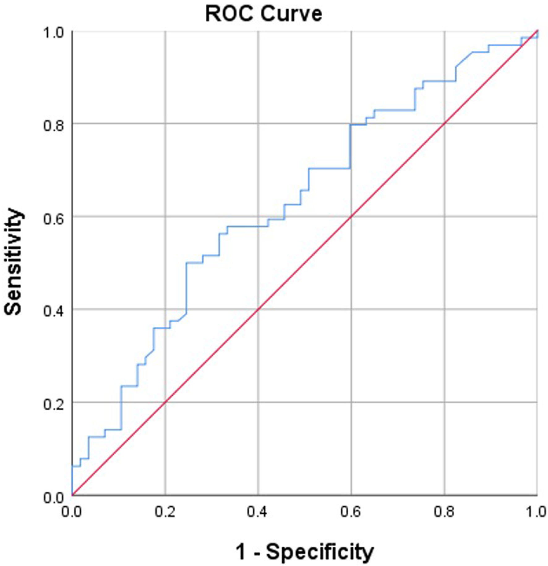 Click for large image | Figure 3. Receiver operating characteristic curve of HPA axis suppression and 8 am cortisol after 3 days of treatment completion (AUC 0.630). HPA: hypothalamic–pituitary–adrenal; AUC: area under the curve. |
Among the 57 patients with biochemical HPA axis suppression at day 3, 18 (31.6%) continued to demonstrate suppression at day 10, while 39 (68.4%) had recovered. Patients with persistent suppression had received glucocorticoids for a longer duration than those who recovered (21.0 ± 7.71 vs. 11.1 ± 5.73 days). Baseline morning cortisol also differed significantly between the groups (P = 0.045) as shown in Table 4. Both glucocorticoid treatment duration and baseline cortisol were significantly associated with persistence of biochemical HPA axis suppression at day 10. Other evaluated clinical and glucocorticoid-related parameters did not demonstrate statistically significant between-group differences.
Click to view | Table 4. Comparison of Baseline Parameters of Study Subjects With and Without HPA Axis Suppression (N = 57) on Day 10 After Completion of Steroid |
Among patients with persistent HPA axis suppression on day 10, given the limited number of persistent-suppression events (n = 18), a parsimonious logistic regression model was constructed using only treatment duration and tapering. Longer treatment duration was associated with increased odds of persistent HPA axis suppression (β = 0.451; adjusted OR: 1.570 per additional day, 95% CI: 1.221–2.018; P < 0.001). Tapering was associated with lower odds of persistent suppression (β = −5.246; adjusted OR: 0.0053, 95% CI: 0.00013–0.207; P = 0.005). However, because of the small number of events and sparse observations, particularly among tapered patients with persistent suppression, these estimates should be considered exploratory and interpreted cautiously. By day 31, all patients had HPA axis recovery.
| Discussion | ▴Top |
Neither the route of steroid administration, cumulative dose, total duration of therapy, nor the underlying disease reliably predict the absence of HPA axis suppression; however, higher doses and prolonged use are associated with the greatest risk [4]. In our study, we evaluated 121 patients who had received varying doses of glucocorticoids for durations ranging from 5 to 45 days, with cumulative doses between 100 and 6,250 mg, to assess their impact on the HPA axis. In our study, HPA axis suppression was evaluated after 3 days of completion of steroid as compared to previous studies that tested HPA axis immediately after completion of steroid therapy. This was done in order to increase the specificity of suppression of HPA axis as the duration of action of long-acting steroids is 72 h. Our study evaluated the integrity of the HPA axis by the low-dose ACTH stimulation test that is the 1-µg ACTH test [21]. The gold standard test to diagnose central adrenal insufficiency is insulin tolerance test (ITT), but this can be unsafe to carry out in patients with history of cardiac disease or seizures [23]. Also, special precautions have to be followed while testing children or the elderly population. The 250-µg ACTH stimulation test assesses adrenal function but is not the preferred method for evaluating central adrenal insufficiency following short-term steroid therapy, as the supraphysiological ACTH dose may mask subtle deficiencies in cortisol secretion [24]. Dickenstein et al established that 1 µg of ACTH is enough to cause the same adrenal response as 250 µg by testing serum cortisol after 30 min of intravenous diluted ACTH preparation. The 1-µg ACTH test is more sensitive than the standard 250 µg ACTH test in detecting central adrenal insufficiency and it closely follows the insulin tolerance test [25]. Additionally, 1–24 ACTH is fully stable in 5-µg/mL saline solution for at least 4 months, in both glass and plastic tubes [21].
We found the prevalence of HPA axis suppression after short-term steroid therapy to be 47% (57/121) on day 3 after completion of therapy, which is comparable to prior studies. Henzen et al studied 75 patients who received the equivalent of at least 25 mg of prednisone daily for 5 to 30 days and found that 45% of patients exhibited HPA axis suppression 2 days after completing therapy [15]. Schuetz et al reported HPA axis suppression in 78% of patients 2 days after and in 33% of patients 7 days after steroid therapy among individuals treated for acute exacerbation of COPD [26]. All patients in our study had HPA axis recovery by 31 days after completion of steroid therapy. In the study by Henzen et al, all but two patients showed recovery of the HPA axis by 14 days after completing steroid therapy [15]. Most of the studies showed recovery of HPA axis within 1 month, with one study of 64 patients by Einaudi et al, showing recovery of HPA axis in all patients by 10 weeks after completion of steroid therapy [27]. All patients with biochemically suppressed HPA axis were initiated on steroid replacement regimen, which could be the reason for a longer duration of 10 weeks that is required for HPA axis recovery. No risk factors for suppression of the HPA axis after the short-term therapy have been identified in previous studies. In our study, we found that the study subjects with HPA axis suppression were significantly younger than those who did not have HPA axis suppression on day 3 after completion of therapy (adjusted OR: 0.963 per year, 95% CI: 0.934–0.993; P = 0.015). In a previous study by Sagar et al, among rheumatology patients receiving long-term steroid therapy, there was no significant association in the age of the patient and HPA axis suppression [20]. However, no explanation can be offered at present as to why younger subjects had more HPA axis suppression.
A study by Ahmad et al argued against tapering of steroids for a short course of steroid therapy and they found recovery of the HPA axis in all patients even after discontinuation of steroid therapy without tapering [28]. General consensus advises against tapering of steroids if the duration of steroid intake is less than 3 weeks [19, 29]. However, in our study, an apparent association between glucocorticoid tapering and a lower frequency of biochemical HPA axis suppression was observed in the unadjusted analysis. However, this finding requires cautious interpretation. Tapering was not randomly assigned and was determined by treating physicians according to clinical circumstances. Consequently, the decision to taper may have been influenced by treatment duration, cumulative glucocorticoid exposure, underlying diagnosis, disease severity, clinical response, and individual prescribing practices, thereby introducing the possibility of confounding by indication.
After revision of the multivariable model to include a more parsimonious set of clinically relevant covariates, the association between tapering and HPA axis suppression was attenuated and no longer reached statistical significance (adjusted OR: 0.448, 95% CI: 0.187–1.075; P = 0.072). Therefore, although the direction of the association suggests a lower frequency of biochemical suppression among patients who underwent tapering, the present data do not demonstrate that tapering itself prevents HPA axis suppression. Prospective studies in which tapering strategies are predefined or randomized would be required to determine whether tapering independently influences HPA axis recovery or the risk of biochemical suppression.
Our study demonstrated that subjects administered less than 500 mg of cumulative steroid dose had significantly more HPA axis suppression on day 3 after steroid completion as compared to subjects administered more than 500 mg of cumulative steroid dose. However, this significance was not present after logistic regression analysis. Hence, the observed association between cumulative dose and HPA axis suppression should be interpreted cautiously. We observed that higher doses of steroids probably had less HPA axis suppression in our study, as higher doses of steroids were tapered before stopping. A tapering strategy may have confounded the relationship between the cumulative dose and HPA axis suppression and that larger studies are required to evaluate their independent and interactive effects. Prior studies with short-term steroid therapy did not reveal any association between the dose of steroid and HPA axis suppression [30]. Meta-analysis of studies including both short-term and long-term steroid intake revealed an increased risk of HPA axis suppression with an increasing dose from 2.4% in low doses to 21.5% in high doses [4]. However, this increased risk of HPA axis suppression with increasing dose of steroid was not demonstrated with a steroid intake of short duration in our study.
In the present study, patients with persistent biochemical HPA axis suppression at day 10 had received glucocorticoid therapy for a longer duration than those who had recovered (21.0 ± 7.71 vs. 11.1 ± 5.73 days). Baseline morning serum cortisol also differed significantly between these groups (P = 0.045). These observations suggest that longer glucocorticoid exposure may be associated with biochemical HPA axis suppression following glucocorticoid withdrawal. These findings, however, should be interpreted cautiously. The analysis involved a relatively small subgroup of patients with persistent suppression, and the observed associations do not establish that treatment duration or baseline cortisol independently predicts delayed recovery. Treatment duration may also be related to the underlying disease, glucocorticoid regimen, cumulative exposure, and clinical decisions regarding treatment discontinuation. Larger prospective studies with serial assessment of HPA axis function are needed to identify reliable predictors of the time to HPA axis recovery following glucocorticoid therapy. Prior studies with short-term therapy did not have a significant correlation with the duration of steroid intake [15, 30]. Our study had no significant difference in study subjects with or without HPA axis suppression based on the type of steroid prescribed or the indication for which the steroid was prescribed.
However, our study was a single-center prospective cohort study involving participants recruited from selected outpatient departments; therefore, selection bias or attrition bias cannot be excluded and may limit generalizability.
The strength of our study includes a well-designed study protocol and adequate sample size. We have adhered strictly to the protocol and have given an accurate representation of the suppression of HPA axis as well as the temporal pattern of recovery of HPA axis. In addition, we have suggested few clinical predictors for the development of HPA axis suppression.
Conclusions
Biochemical HPA axis suppression after short-course systemic glucocorticoid therapy was common but transient, with recovery occurring in all patients within 1 month of steroid discontinuation. Total cumulative dose of steroids, total duration of steroid therapy or type of steroid used did not predict HPA axis suppression after a short course of steroid therapy. Baseline morning cortisol may help identify patients at greater risk of subsequent biochemical suppression. Although a lower frequency of HPA axis suppression was observed among patients who underwent glucocorticoid tapering, this association was not statistically significant after multivariable adjustment and should not be interpreted as evidence of a causal protective effect. Further prospective studies are required to determine whether specific glucocorticoid discontinuation or tapering strategies influence HPA axis suppression and recovery.
Acknowledgments
The authors express gratitude to the participants in the study. They also express gratitude to the Endocrine Society of India for funding the research.
Financial Disclosure
The authors received the Dissertation Grant from Endocrine Society of India (ESI Trainee Grant).
Conflict of Interest
The authors declare that they have no financial or non-financial conflict of interest to report.
Informed Consent
Informed consent was obtained from all individual participants included in the study.
Author Contributions
Conception and design of the research, JB and PJ. Assisted in data collection, PJ, SS, and JB. Analysis and interpretation of the data, PJ, SR, JS, and BK. Statistical analysis, JB, PJ, and SM. Writing of the manuscript, PJ, JB, and SM. Critical revision of the manuscript, AS, JS, SR, and BK. All authors have read and approved the final manuscript submitted.
Data Availability
Any inquiries regarding the availability of the supporting data from this study should be directed to the corresponding author.
Abbreviations
AUC: area under the curve; ACTH: adrenocorticotrophic hormone; AIHA: auto-immune hemolytic anemia; BMI: body mass index; CI: confidence interval; CNS: central nervous system; COPD: chronic obstructive pulmonary disease; ECLIA: electrochemiluminescence immunoassay; HPA: hypothalamic–pituitary–adrenal; ITT: insulin tolerance test; OR: odds ratio; ROC: receiver operating characteristic; SD: standard deviation; VIFs: variance inflation factors
| References | ▴Top |
- Prete A, Bancos I. Glucocorticoid induced adrenal insufficiency. BMJ. 2021;374:n1380.
doi pubmed - Mebrahtu TF, Morgan AW, Keeley A, Baxter PD, Stewart PM, Pujades-Rodriguez M. Dose dependency of iatrogenic glucocorticoid excess and adrenal insufficiency and mortality: a cohort study in England. J Clin Endocrinol Metab. 2019;104(9):3757-3767.
doi pubmed - Joseph RM, Hunter AL, Ray DW, Dixon WG. Systemic glucocorticoid therapy and adrenal insufficiency in adults: a systematic review. Semin Arthritis Rheum. 2016;46(1):133-141.
doi pubmed - Broersen LH, Pereira AM, Jorgensen JO, Dekkers OM. Adrenal insufficiency in corticosteroids use: systematic review and meta-analysis. J Clin Endocrinol Metab. 2015;100(6):2171-2180.
doi pubmed - Bornstein SR, Allolio B, Arlt W, Barthel A, Don-Wauchope A, Hammer GD, Husebye ES, et al. Diagnosis and treatment of primary adrenal insufficiency: an endocrine society clinical practice guideline. J Clin Endocrinol Metab. 2016;101(2):364-389.
doi pubmed - Rushworth RL, Torpy DJ, Falhammar H. Adrenal crisis. N Engl J Med. 2019;381(9):852-861.
doi pubmed - Smans LC, Van der Valk ES, Hermus AR, Zelissen PM. Incidence of adrenal crisis in patients with adrenal insufficiency. Clin Endocrinol (Oxf). 2016;84(1):17-22.
doi pubmed - Laugesen K, Broersen LHA, Hansen SB, Dekkers OM, Sorensen HT, Jorgensen JOL. Management of endocrine disease: glucocorticoid-induced adrenal insufficiency: replace while we wait for evidence? Eur J Endocrinol. 2021;184(4):R111-R122.
doi pubmed - Crowley RK, Argese N, Tomlinson JW, Stewart PM. Central hypoadrenalism. J Clin Endocrinol Metab. 2014;99(11):4027-4036.
doi pubmed - Paragliola RM, Papi G, Pontecorvi A, Corsello SM. Treatment with synthetic glucocorticoids and the hypothalamus-pituitary-adrenal axis. Int J Mol Sci. 2017;18(10):2201.
doi pubmed - Sacre K, Dehoux M, Chauveheid MP, Chauchard M, Lidove O, Roussel R, Papo T. Pituitary-adrenal function after prolonged glucocorticoid therapy for systemic inflammatory disorders: an observational study. J Clin Endocrinol Metab. 2013;98(8):3199-3205.
doi pubmed - Jamilloux Y, Liozon E, Pugnet G, Nadalon S, Heang Ly K, Dumonteil S, Gondran G, et al. Recovery of adrenal function after long-term glucocorticoid therapy for giant cell arteritis: a cohort study. PLoS One. 2013;8(7):e68713.
doi pubmed - Levic Z, Micic D, Nikolic J, Stojisavljevic N, Sokic D, Jankovic S, Kendereski A, et al. Short-term high dose steroid therapy does not affect the hypothalamic-pituitary-adrenal axis in relapsing multiple sclerosis patients. Clinical assessment by the insulin tolerance test. J Endocrinol Invest. 1996;19(1):30-34.
doi pubmed - Aljebab F, Choonara I, Conroy S. Systematic review of the toxicity of short-course oral corticosteroids in children. Arch Dis Child. 2016;101(4):365-370.
doi pubmed - Henzen C, Suter A, Lerch E, Urbinelli R, Schorno XH, Briner VA. Suppression and recovery of adrenal response after short-term, high-dose glucocorticoid treatment. Lancet. 2000;355(9203):542-545.
doi pubmed - Schuetz P, Leuppi JD, Bingisser R, Bodmer M, Briel M, Drescher T, Duerring U, et al. Prospective analysis of adrenal function in patients with acute exacerbations of COPD: the Reduction in the Use of Corticosteroids in Exacerbated COPD (REDUCE) trial. Eur J Endocrinol. 2015;173(1):19-27.
doi pubmed - Han HS, Park JC, Park SY, Lee KT, Bae SB, Kim HJ, Kim S, et al. A prospective multicenter study evaluating secondary adrenal suppression after antiemetic dexamethasone therapy in cancer patients receiving chemotherapy: a Korean South West Oncology Group Study. Oncologist. 2015;20(12):1432-1439.
doi pubmed - Rensen N, Gemke RJ, van Dalen EC, Rotteveel J, Kaspers GJ. Hypothalamic-pituitary-adrenal (HPA) axis suppression after treatment with glucocorticoid therapy for childhood acute lymphoblastic leukaemia. Cochrane Database Syst Rev. 2017;11(11):CD008727.
doi pubmed - Melmed S, Auchus Rj, Goldfine AB, et al. Williams textbook of endocrinology. 15th ed. Pliladelphia, PA: Elsevier; 2025.
- Sagar R, Mackie S, A WM, Stewart P, Abbas A. Evaluating tertiary adrenal insufficiency in rheumatology patients on long-term systemic glucocorticoid treatment. Clin Endocrinol (Oxf). 2021;94(3):361-370.
doi pubmed - Dickstein G, Shechner C, Nicholson WE, Rosner I, Shen-Orr Z, Adawi F, Lahav M. Adrenocorticotropin stimulation test: effects of basal cortisol level, time of day, and suggested new sensitive low dose test. J Clin Endocrinol Metab. 1991;72(4):773-778.
doi pubmed - Dickstein G, Shechner C. Low dose ACTH test—a word of caution to the word of caution: when and how to use it. J Clin Endocrinol Metab. 1997;82(1):322.
doi pubmed - Erturk E, Jaffe CA, Barkan AL. Evaluation of the integrity of the hypothalamic-pituitary-adrenal axis by insulin hypoglycemia test. J Clin Endocrinol Metab. 1998;83(7):2350-2354.
doi pubmed - Ospina NS, Al Nofal A, Bancos I, Javed A, Benkhadra K, Kapoor E, Lteif AN, et al. ACTH stimulation tests for the diagnosis of adrenal insufficiency: systematic review and meta-analysis. J Clin Endocrinol Metab. 2016;101(2):427-434.
doi pubmed - Rasmuson S, Olsson T, Hagg E. A low dose ACTH test to assess the function of the hypothalamic-pituitary-adrenal axis. Clin Endocrinol (Oxf). 1996;44(2):151-156.
doi pubmed - Schuetz P, Christ-Crain M, Schild U, Suess E, Facompre M, Baty F, Nusbaumer C, et al. Effect of a 14-day course of systemic corticosteroids on the hypothalamic-pituitary-adrenal-axis in patients with acute exacerbation of chronic obstructive pulmonary disease. BMC Pulm Med. 2008;8:1.
doi pubmed - Einaudi S, Bertorello N, Masera N, Farinasso L, Barisone E, Rizzari C, Corrias A, et al. Adrenal axis function after high-dose steroid therapy for childhood acute lymphoblastic leukemia. Pediatr Blood Cancer. 2008;50(3):537-541.
doi pubmed - Ahmad N, Abosoudah IF, Sobaihi MM, Algiraigri AH, Roujouleh F, Ghurab F, Chanoine JP. Adrenal function following acute discontinuation of glucocorticoids in children with acute lymphocytic leukemia: A prospective study. Pediatr Hematol Oncol. 2019;36(7):422-431.
doi pubmed - LaRochelle GE, Jr., LaRochelle AG, Ratner RE, Borenstein DG. Recovery of the hypothalamic-pituitary-adrenal (HPA) axis in patients with rheumatic diseases receiving low-dose prednisone. Am J Med. 1993;95(3):258-264.
doi pubmed - Spiegel RJ, Vigersky RA, Oliff AI, Echelberger CK, Bruton J, Poplack DG. Adrenal suppression after short-term corticosteroid therapy. Lancet. 1979;1(8117):630-633.
doi pubmed
This article is distributed under the terms of the Creative Commons Attribution 4.0 International License (CC BY 4.0), which permits unrestricted use, distribution, and reproduction in any medium, including commercial use, provided the original work is properly cited.
Journal of Endocrinology and Metabolism is published by Elmer Press Inc.